


Content of
Respiratory System Physiology & Anatomy PowerPoint Presentation
Slide 1: Respiratory System (Cover Slide)
.PNG)
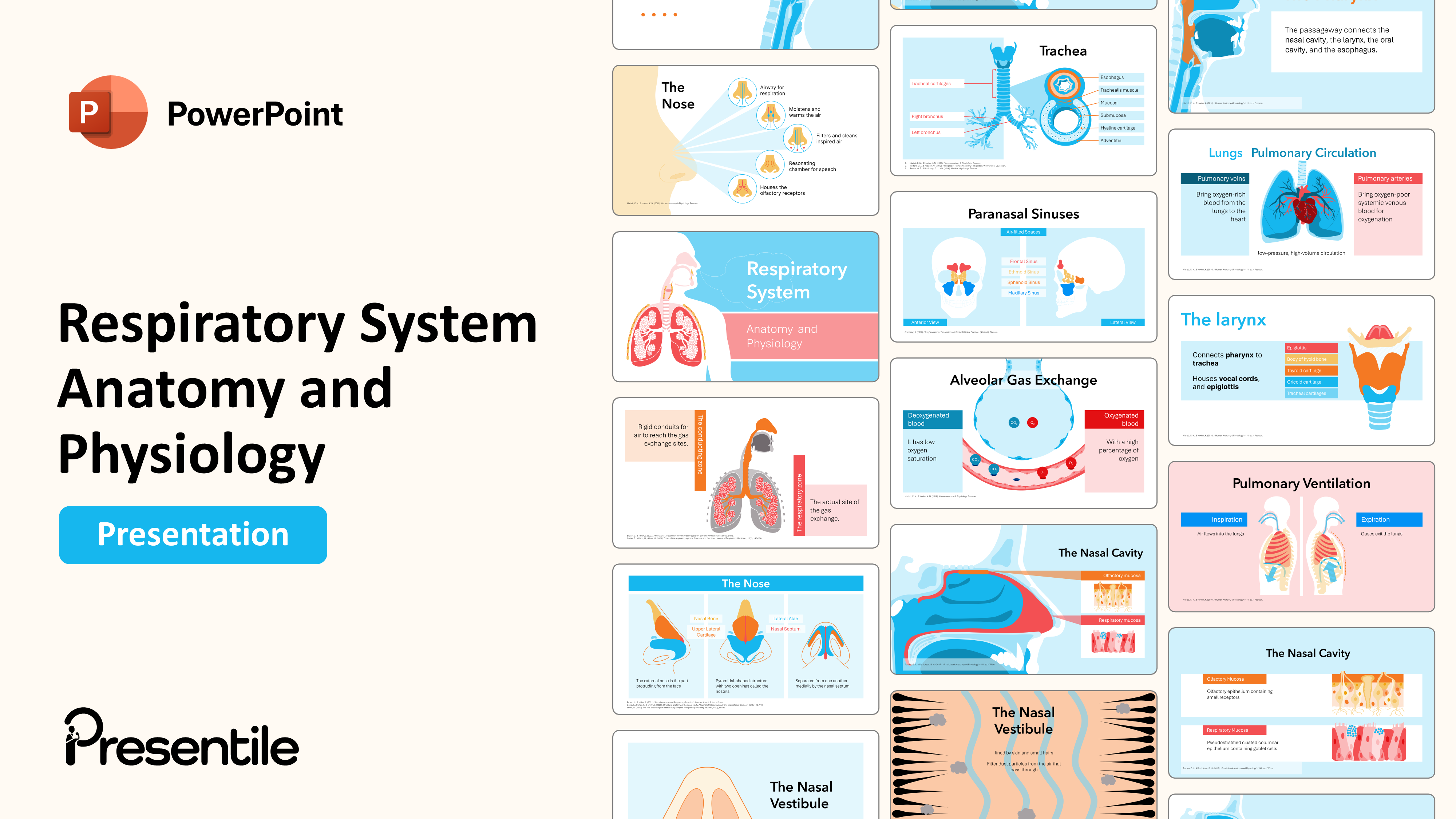
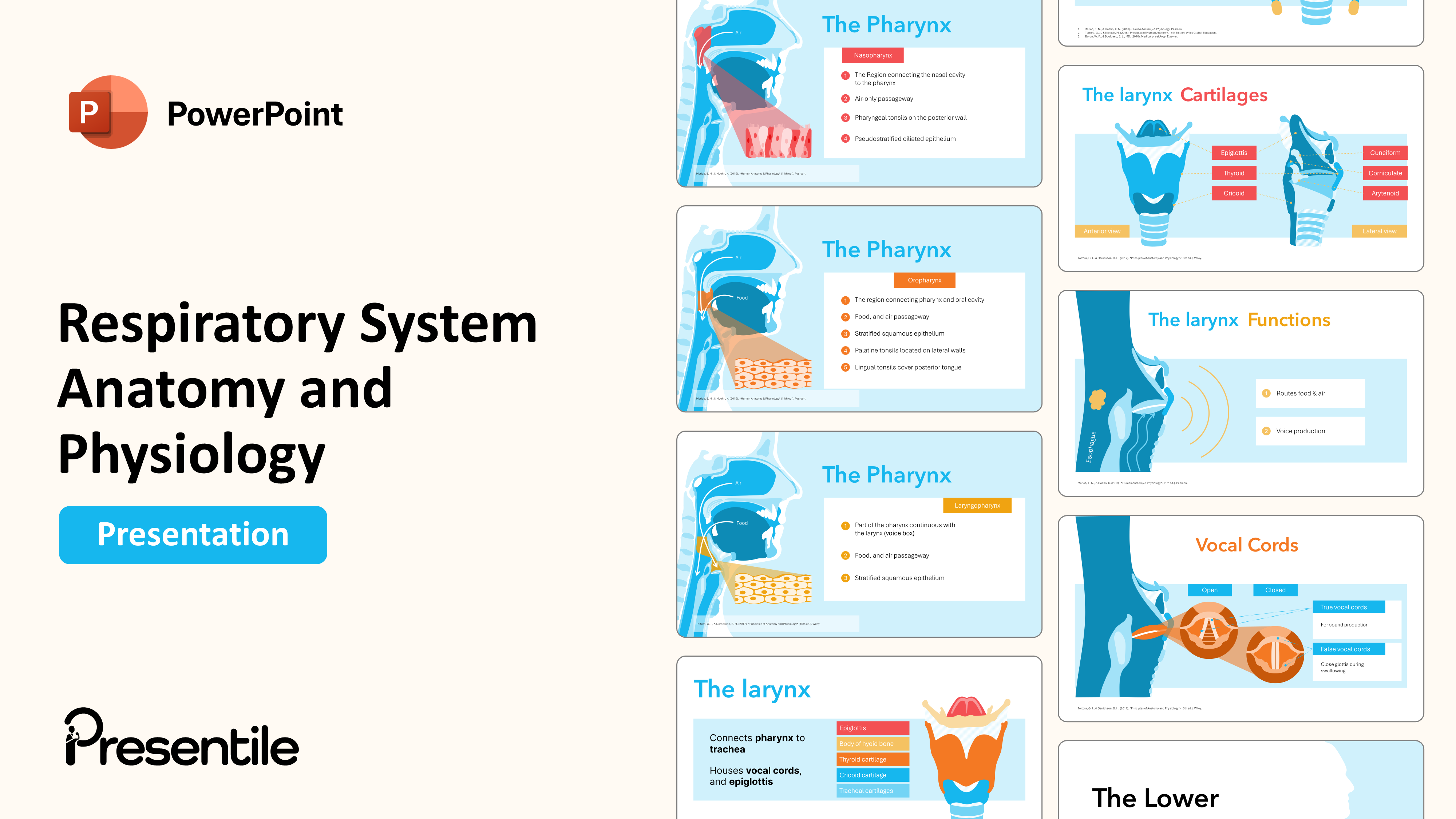
- This professional medical PowerPoint template is an ideal title slide for a presentation on the human respiratory system.
- Featuring a clear, color-blocked layout with a detailed anatomical illustration of the lungs and upper airway, this professional slide sets a clean and educational tone.
- Its editable design is perfect for educators, students, and healthcare professionals needing a strong and impactful cover slide for their clinical presentation on anatomy and physiology.
Slide 2: Respiratory System Anatomy (Section Slide)
.PNG)
- This professional medical PowerPoint template provides a clean and Section slide for discussing the anatomy of the respiratory system.
- The layout features a prominent, stylized illustration of the lungs on a light blue background, complemented by a clear title and subtitle on the left.
Slide 3: Upper respiratory tract

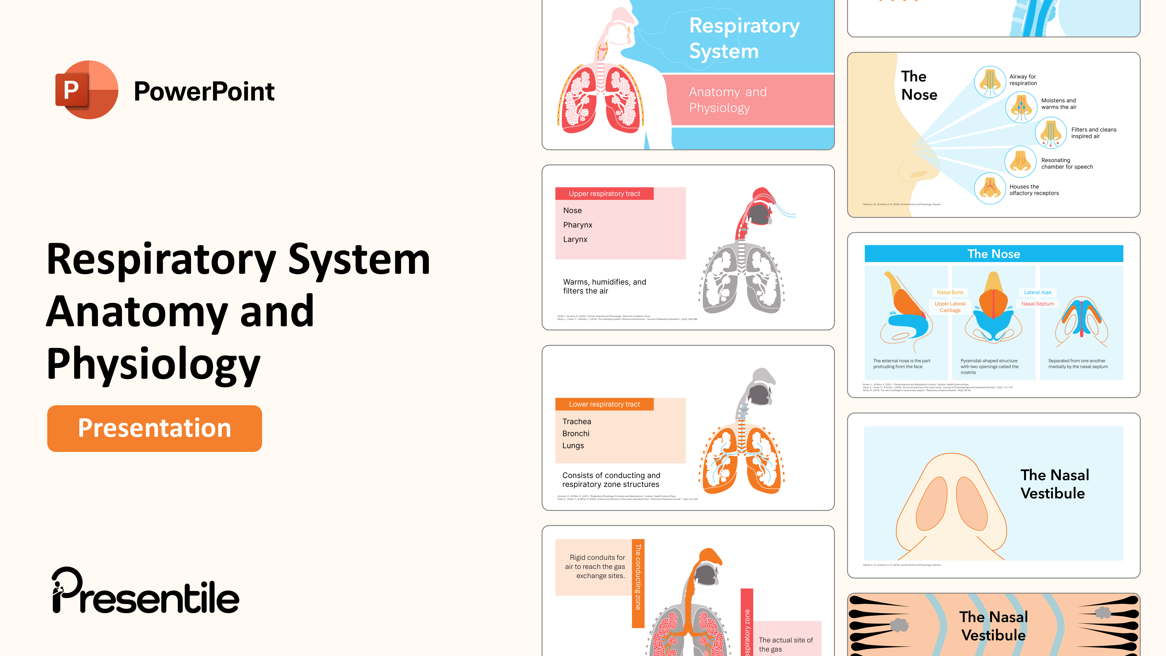
- This professional medical PowerPoint template provides a clear and organized overview of the Upper respiratory tract.
- The slide lists the key components—Nose, Pharynx, and Larynx—and their collective function to Warms, humidifies, and filters the air.
- A stylized anatomical illustration to the right visually complements the text, making this editable layout perfect for any clinical presentation or educational lecture on the respiratory system.
Slide 4: Lower respiratory tract
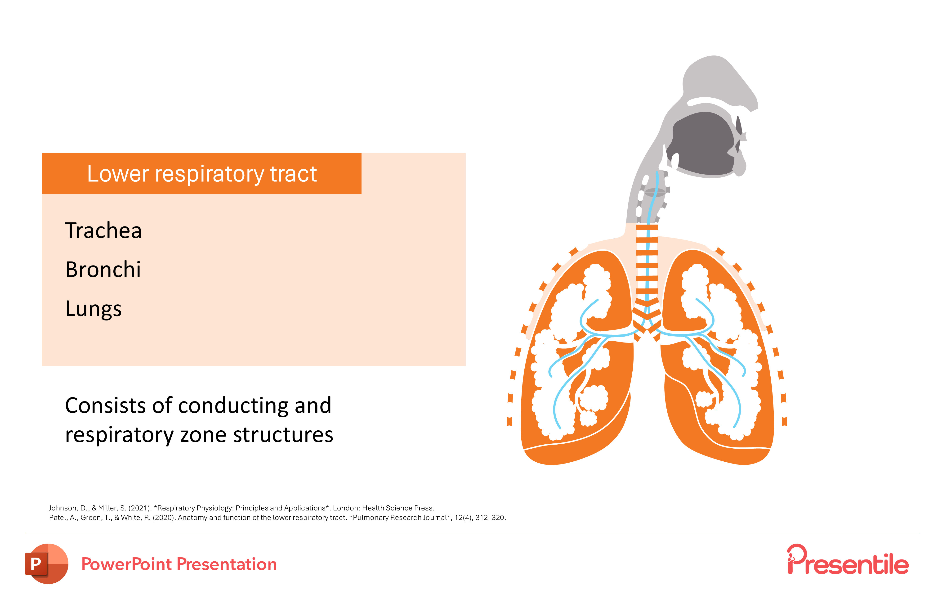
- This professional medical PowerPoint template provides a clear and organized overview of the Lower respiratory tract.
- The slide lists its key anatomical components—Trachea, Bronchi, and Lungs—and notes that it Consists of conducting and respiratory zone structures.
- A stylized illustration on the right visually highlights this section of the respiratory system, making this editable layout perfect for any clinical presentation or educational lecture on human anatomy.
Slide 5: Functional zones of the respiratory system

- This professional medical PowerPoint template expertly explains the functional zones of the respiratory system.
- Using a clear, color-coded anatomical illustration, the slide distinguishes between the conducting zone—described as "Rigid conduits for air to reach the gas exchange sites"—and the respiratory zone, which is "The actual site of the gas exchange."
Slide 6: The Upper Respiratory Tract ( Section Slide)
.PNG)
- This professional medical PowerPoint template provides a clean and section slide for introducing the anatomy of The Upper Respiratory Tract.
- The layout features a prominent, stylized illustration of the nasal and oral cavities on the right, providing a clear visual anchor.
Slide 7: The Nose

- This professional medical PowerPoint template offers a detailed and organized breakdown of the key functions of The Nose.
- The slide's visual layout features a stylized illustration on the left, with five corresponding diagrams on the right that explain its roles, including its function as an Airway for respiration and how it Moistens and warms the air.
Slide 8: The Nose cont.
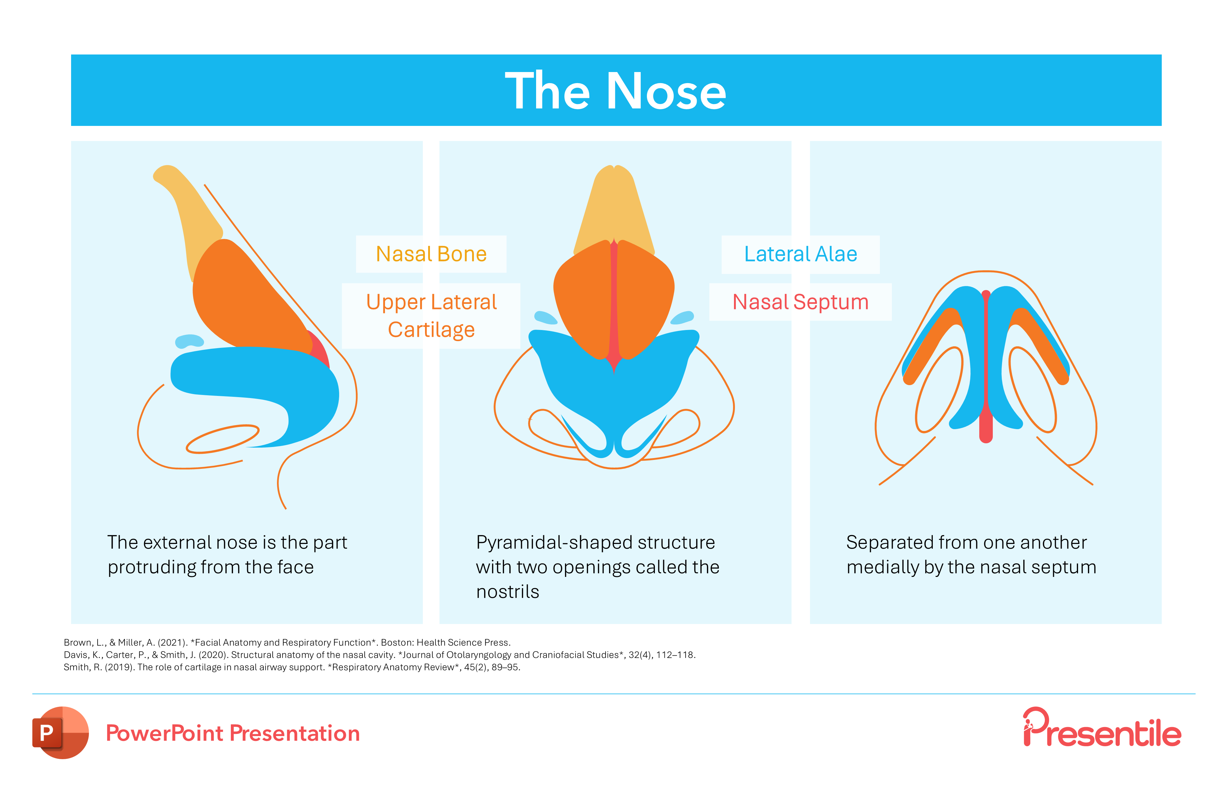
- This professional medical PowerPoint template offers a detailed anatomical look at the external nose with a multi-panel layout.
- The slide presents three distinct views—profile, frontal, and cross-section—to illustrate key structures such as the Nasal Bone, Upper Lateral Cartilage, and Nasal Septum. With its clean design and clearly labeled diagrams.
Slide 9: The Nasal Vestibule (Section Slide)

- This professional medical PowerPoint template offers a clean and focused slide for introducing the anatomy of The Nasal Vestibule.
- The simple layout features a stylized illustration of the nostrils on the left, providing a clear visual anchor, while the right side is dedicated to a bold title.
Slide 10: The Nasal Vestibule
.PNG)
- This professional medical PowerPoint template titled The Nasal Vestibule provides a clear and unique visual explanation of its function.
- The slide uses a stylized graphic to show how the vestibule, lined by skin and small hairs, serves to Filter dust particles from the air that pass through.
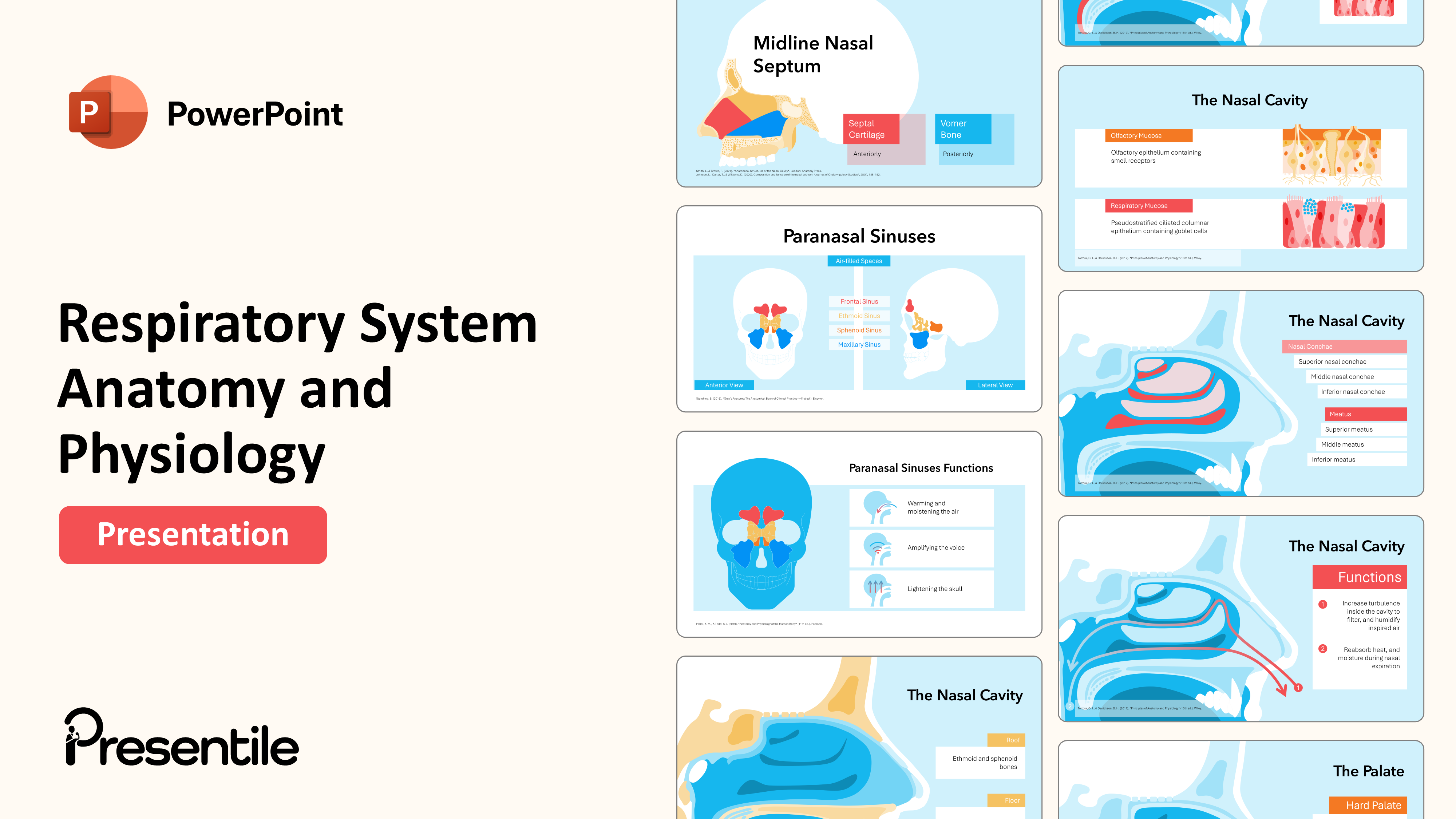
Slide 11: Midline Nasal Septum
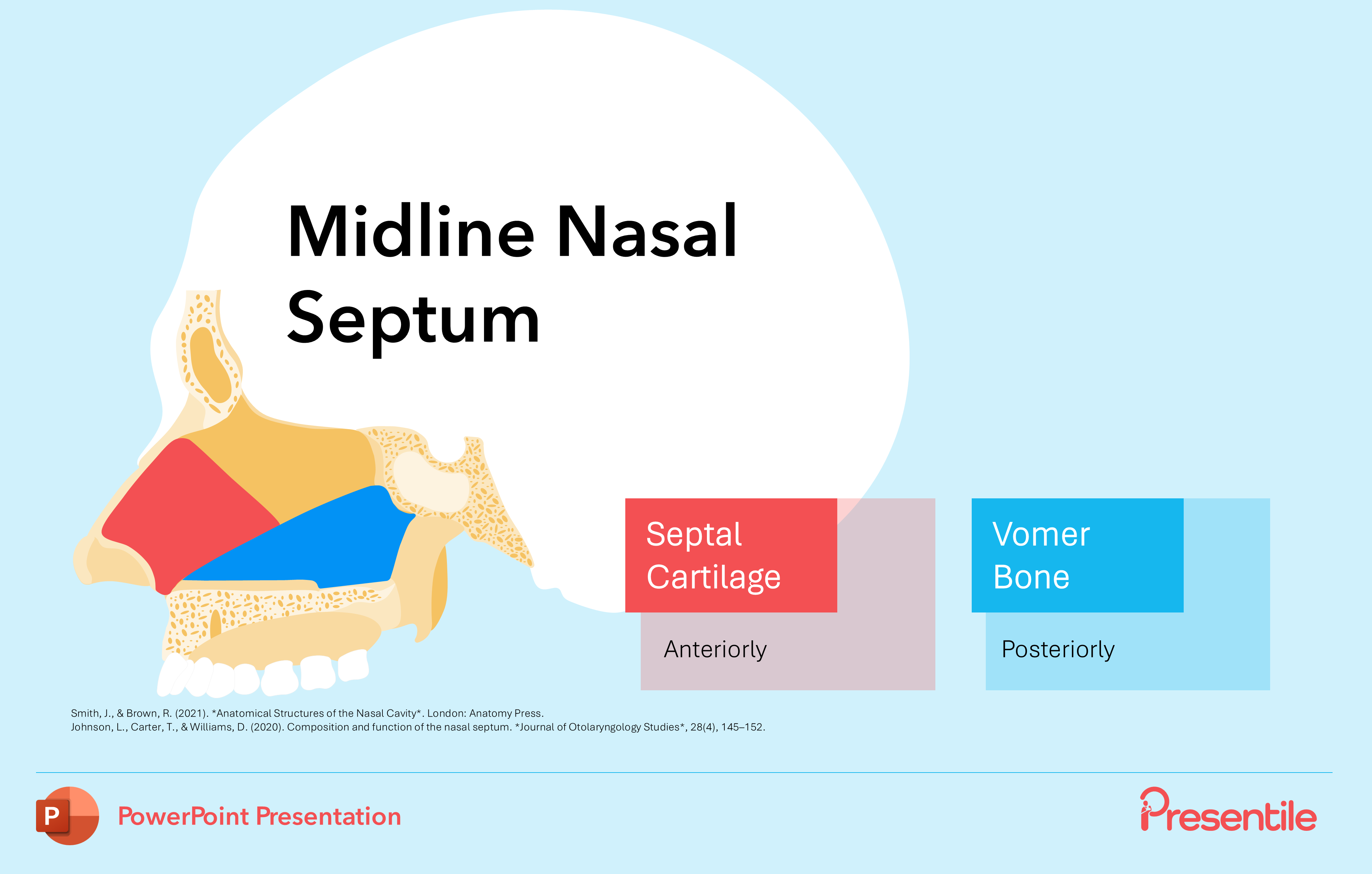
- This professional medical PowerPoint template provides a focused look at the anatomy of the Midline Nasal Septum.
- The slide’s clear and editable layout features a stylized diagram that visually breaks down the two main components: the Septal Cartilage, which is located anteriorly, and the Vomer Bone, which is located posteriorly.
Slide 12: Paranasal Sinuses
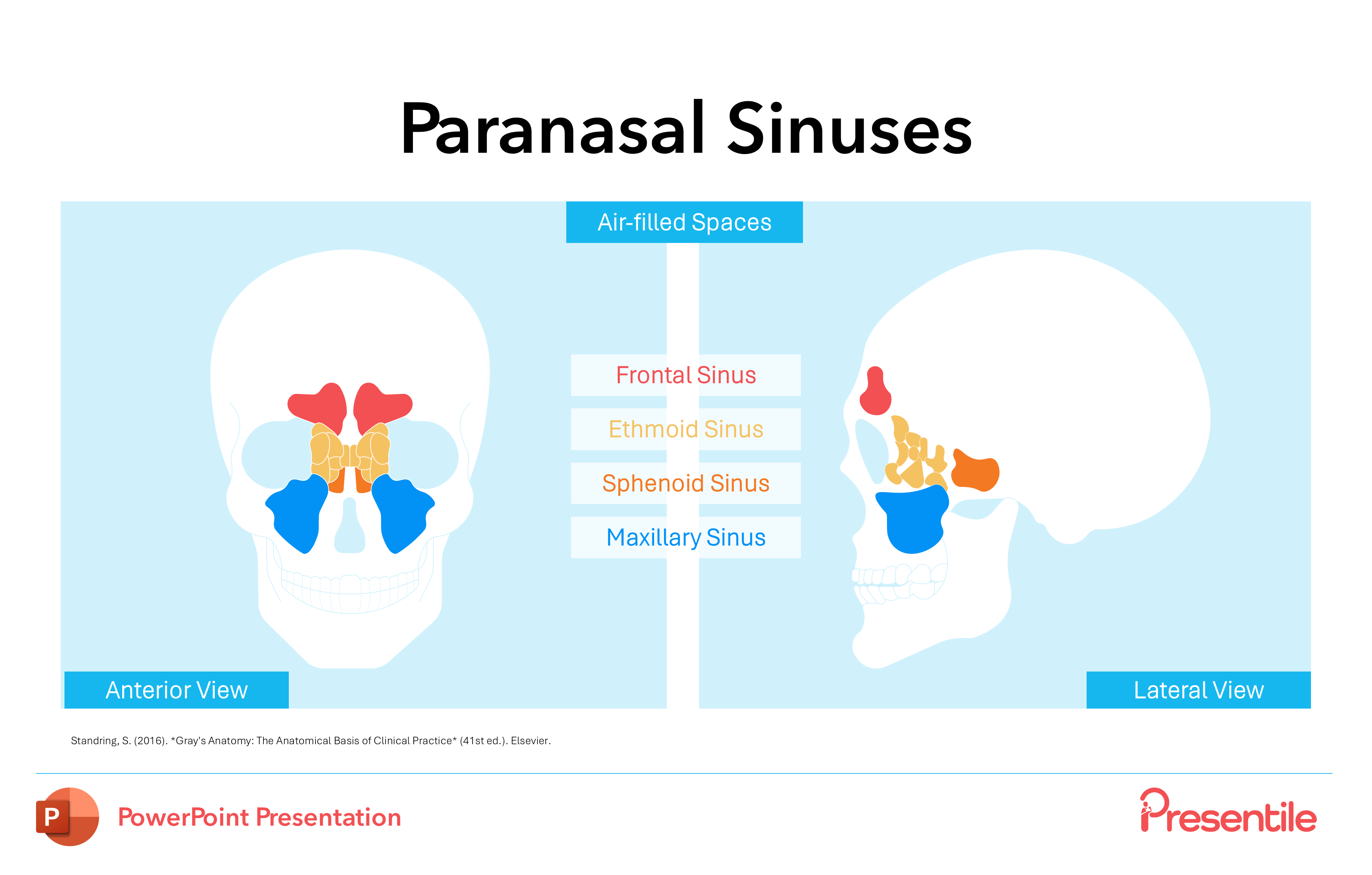
- This professional medical PowerPoint template offers a clear and detailed look at the Paranasal Sinuses.
- The slide’s clean, two-panel layout presents both an Anterior View and a Lateral View of the skull, which highlights the location of the four major air-filled spaces: the Frontal, Ethmoid, Sphenoid, and Maxillary Sinuses.
Slide 13: Paranasal Sinuses Functions
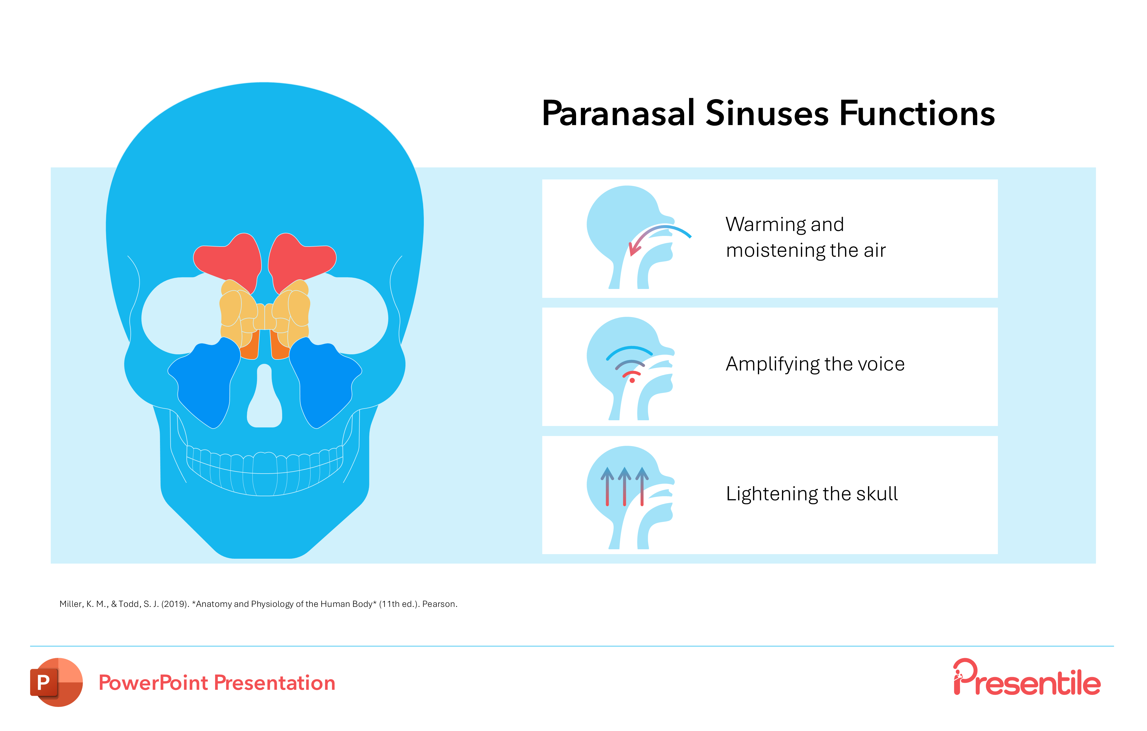
- Provides a clear, functional overview of the Paranasal Sinuses.
- The slide’s clean layout features a large anatomical illustration on the left, while the right side is dedicated to a list of three key physiological functions: Warming and moistening the air, Amplifying the voice, and Lightening the skull.
Slide 14: The Nasal Cavity
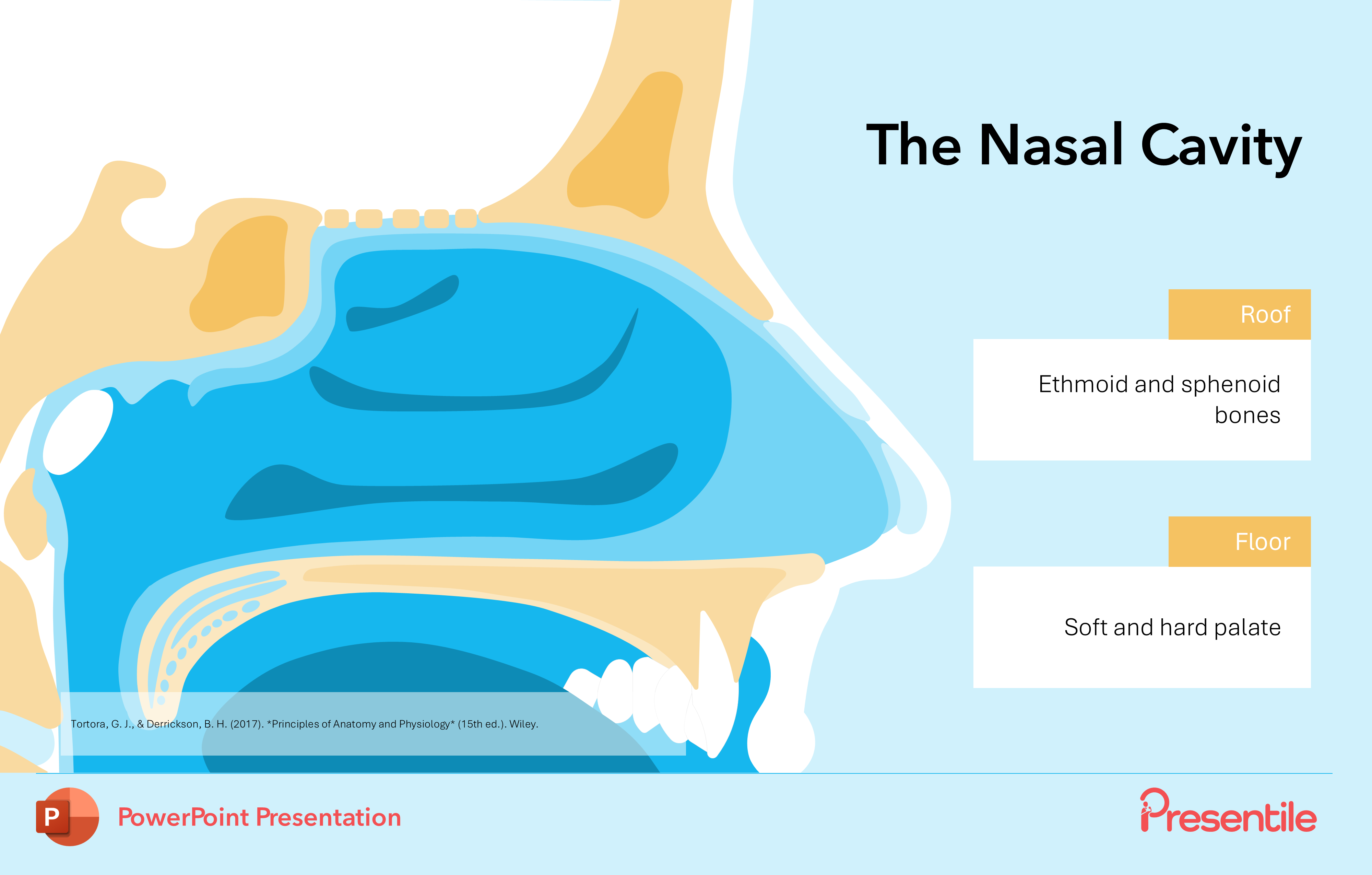
- This initial slide provides a clear and detailed anatomical overview of the nasal cavity.
- It features a high-quality, illustrative diagram that clearly labels the key structural components of the nasal cavity's roof and floor, specifically identifying the ethmoid and sphenoid bones, as well as the soft and hard palates.
- The roof of the nasal cavity is formed by the ethmoid and sphenoid bones, which provide structural support and house important anatomical features,
- In contrast, the floor of the nasal cavity is formed by the hard palate and the soft palate.
Slide 15: The Nasal Cavity Cont.
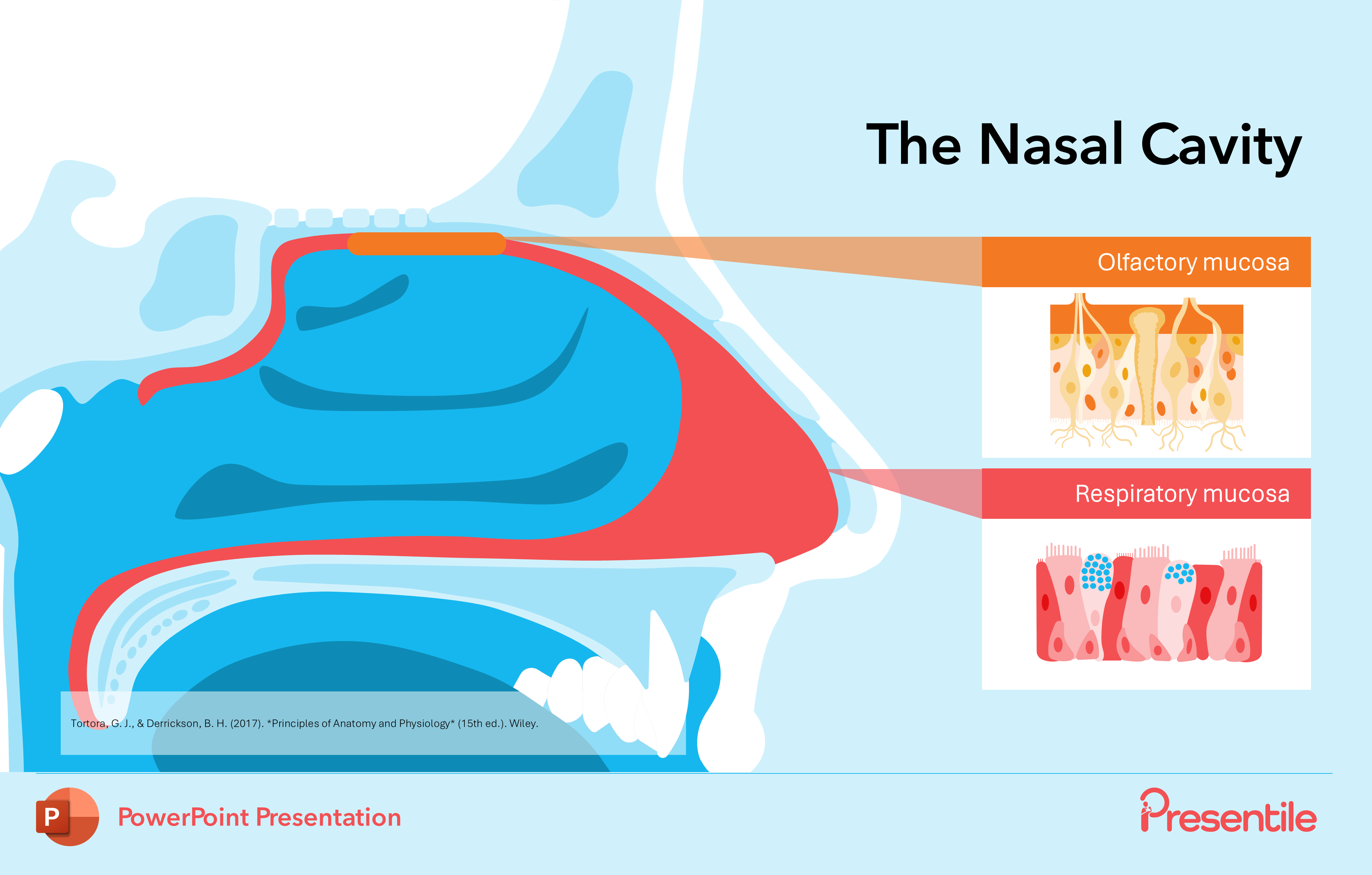
- This slide builds upon the anatomical overview by focusing on the crucial tissues that line the nasal cavity.
- It features a detailed, side-by-side comparison of the olfactory mucosa and the respiratory mucosa, using zoomed-in illustrations to highlight their distinct cellular structures.
- The nasal cavity is lined by two distinct types of mucosa, as depicted in these diagrams: the olfactory mucosa and the respiratory mucosa.
Slide 16: Olfactory Mucosa Vs Respiratory Mucosa
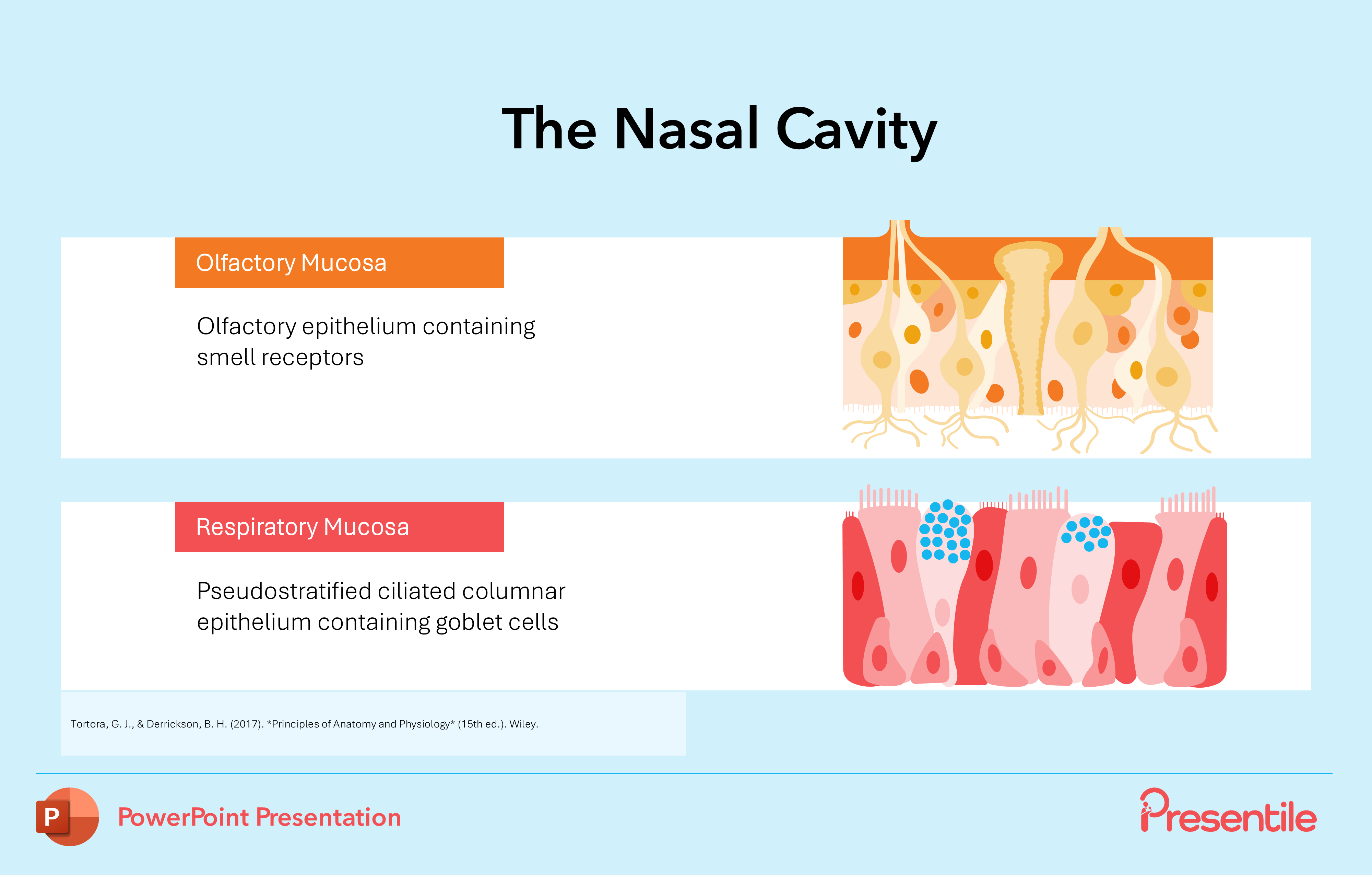
- This slide provides a more in-depth histological view of the nasal mucosa, detailing the specific cellular composition of each tissue type.
- It breaks down the olfactory mucosa, highlighting the olfactory epithelium and its smell receptors, and contrasts it with the respiratory mucosa, detailing the pseudostratified ciliated columnar epithelium and the presence of goblet cells.
Slide 17: The Structure of The Nasal Conchae
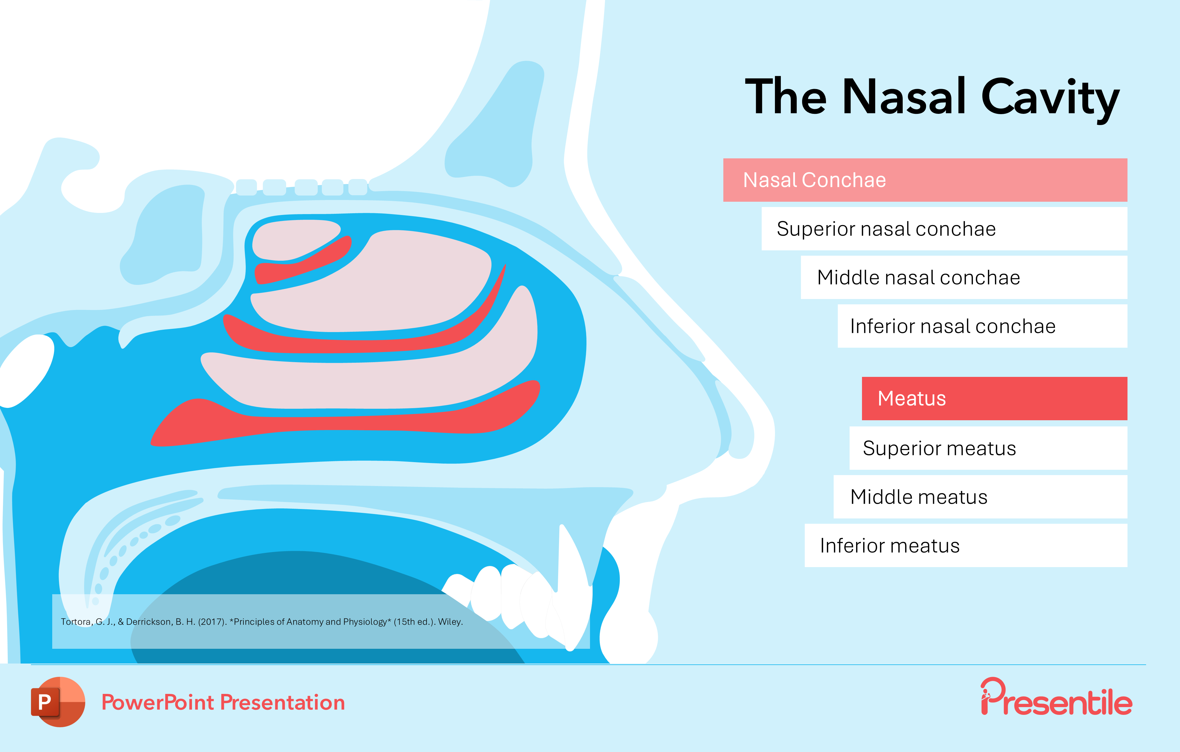
- This slide deepens the anatomical exploration by detailing the structure of the nasal conchae and meatuses.
- The clear, color-coded diagram distinguishes and labels the superior, middle, and inferior conchae and their corresponding meatuses.
Slide 18: Key Functions of The Nasal Cavity

- This slide transitions from anatomy to physiology by detailing the key functions of the nasal cavity.
- Using clear, numbered points and a dynamic diagram with arrows illustrating airflow, it explains how the nasal passages increase air turbulence to efficiently filter, warm, and humidify inspired air.
- It also covers the vital role of the nasal cavity in reabsorbing heat and moisture during expiration, providing a comprehensive overview of its role in respiratory function.
Slide 19: The Palate
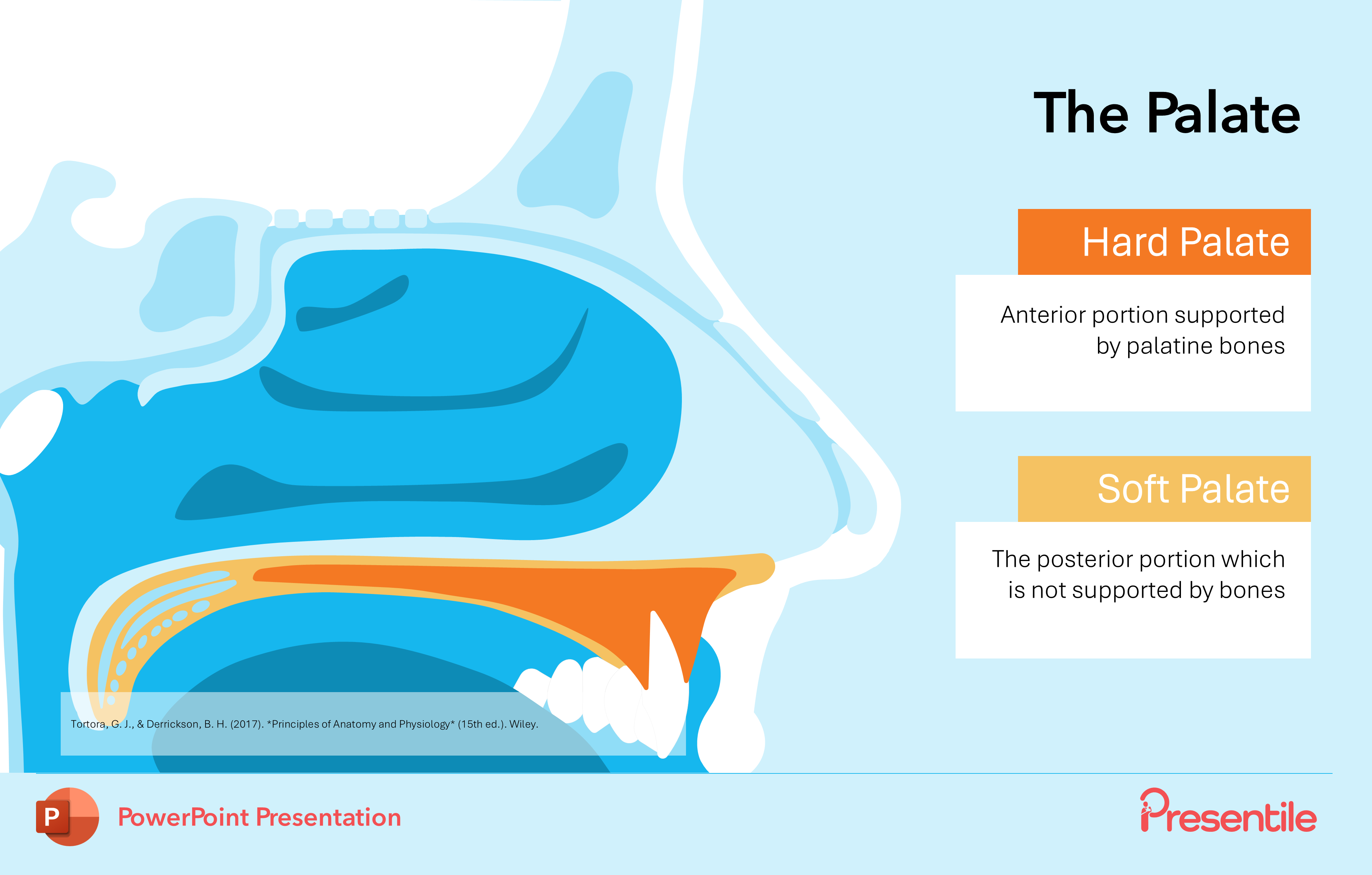
- This slide hones in on the structure of the palate, a key component of the nasal cavity's floor.
- It uses a clear visual to differentiate and label the anterior hard palate and the posterior soft palate.
- The key anatomical distinction—that the hard palate is supported by bone while the soft palate is not—providing a fundamental understanding of their respective structures and locations.
Slide 20: The Uvula
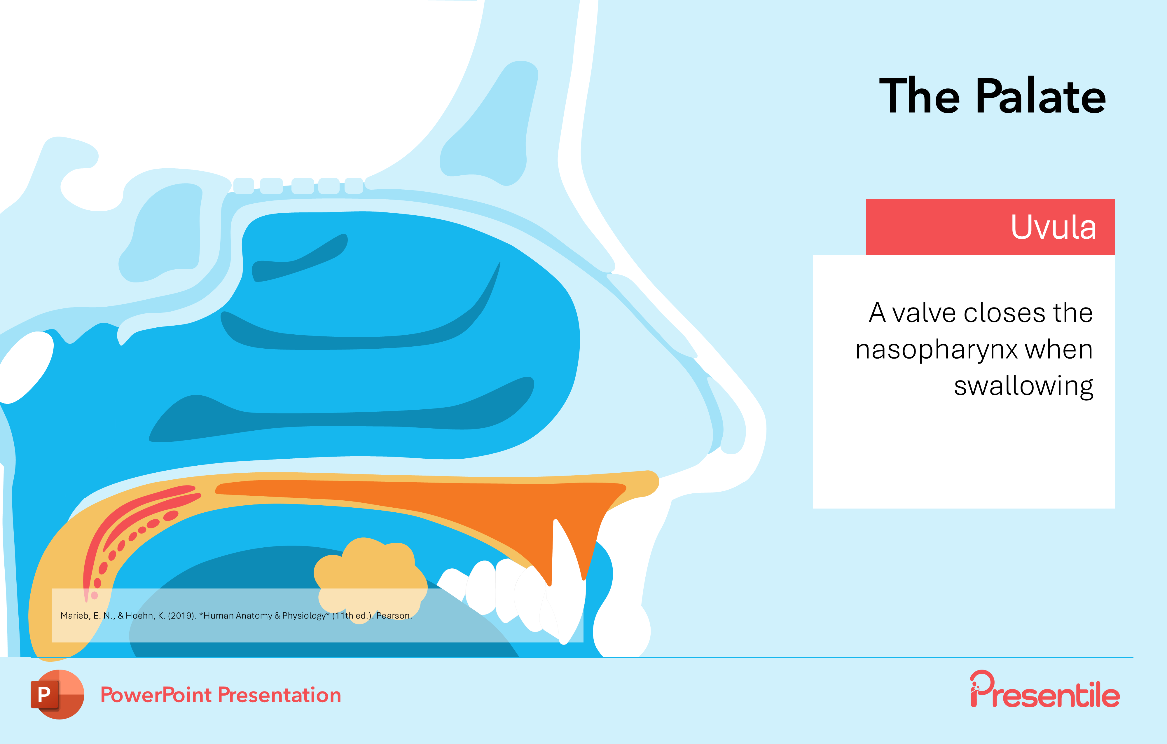
- This slide specifically focuses on the uvula, a key part of the soft palate. It visually isolates the uvula and concisely describes its critical function: acting as a valve that closes off the nasopharynx during swallowing. This detail is essential for a complete understanding of how the nasal cavity is protected from food and liquid during the digestive process.
Slide 21: The Pharynx
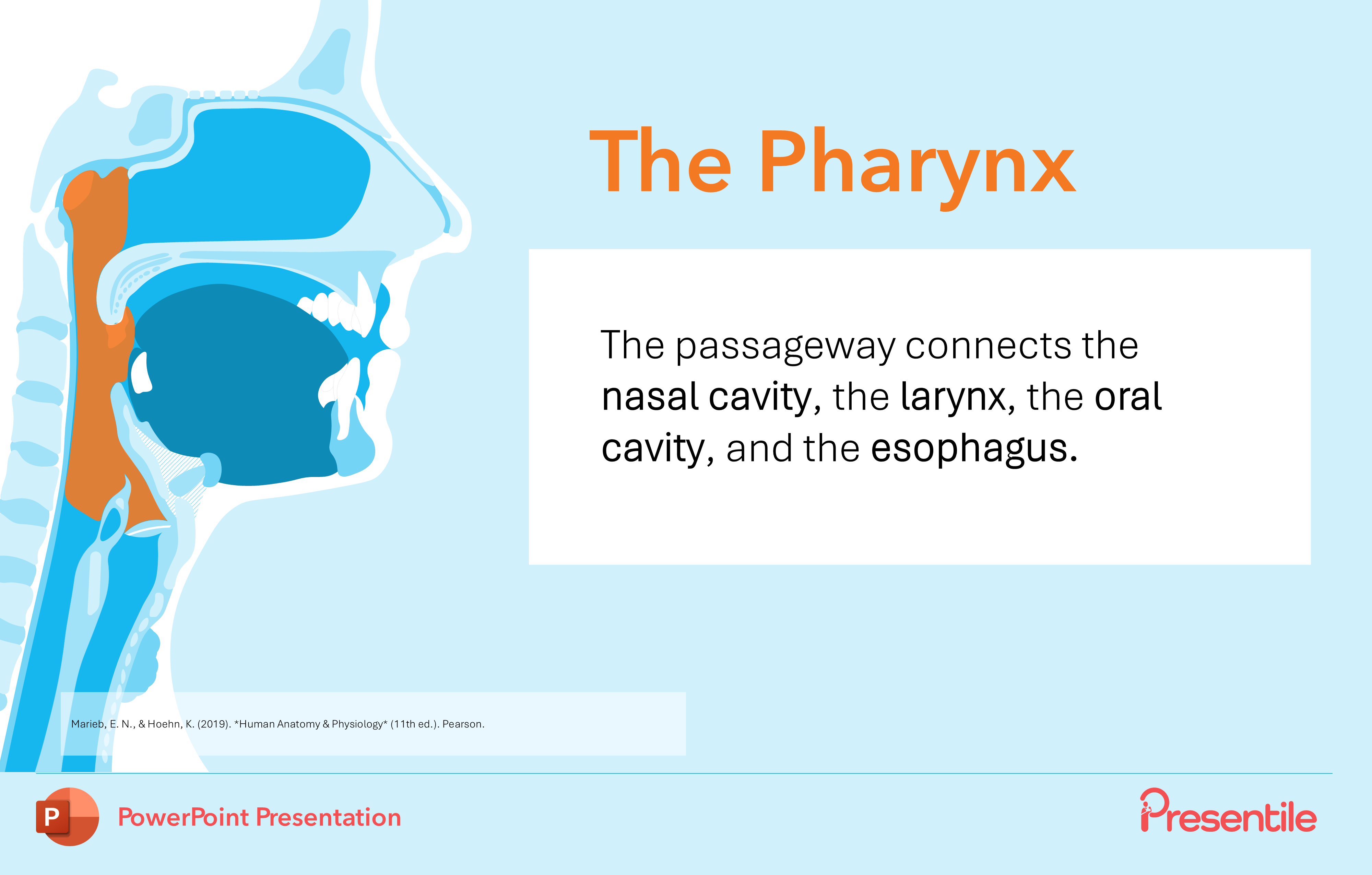
- This slide introduces the pharynx, a vital anatomical structure that serves as a passageway connecting multiple key areas of the head and neck.
- The clear diagram visually identifies the pharynx as the intersection between the nasal cavity, oral cavity, larynx, and esophagus.
Slide 22: The Nasopharynx
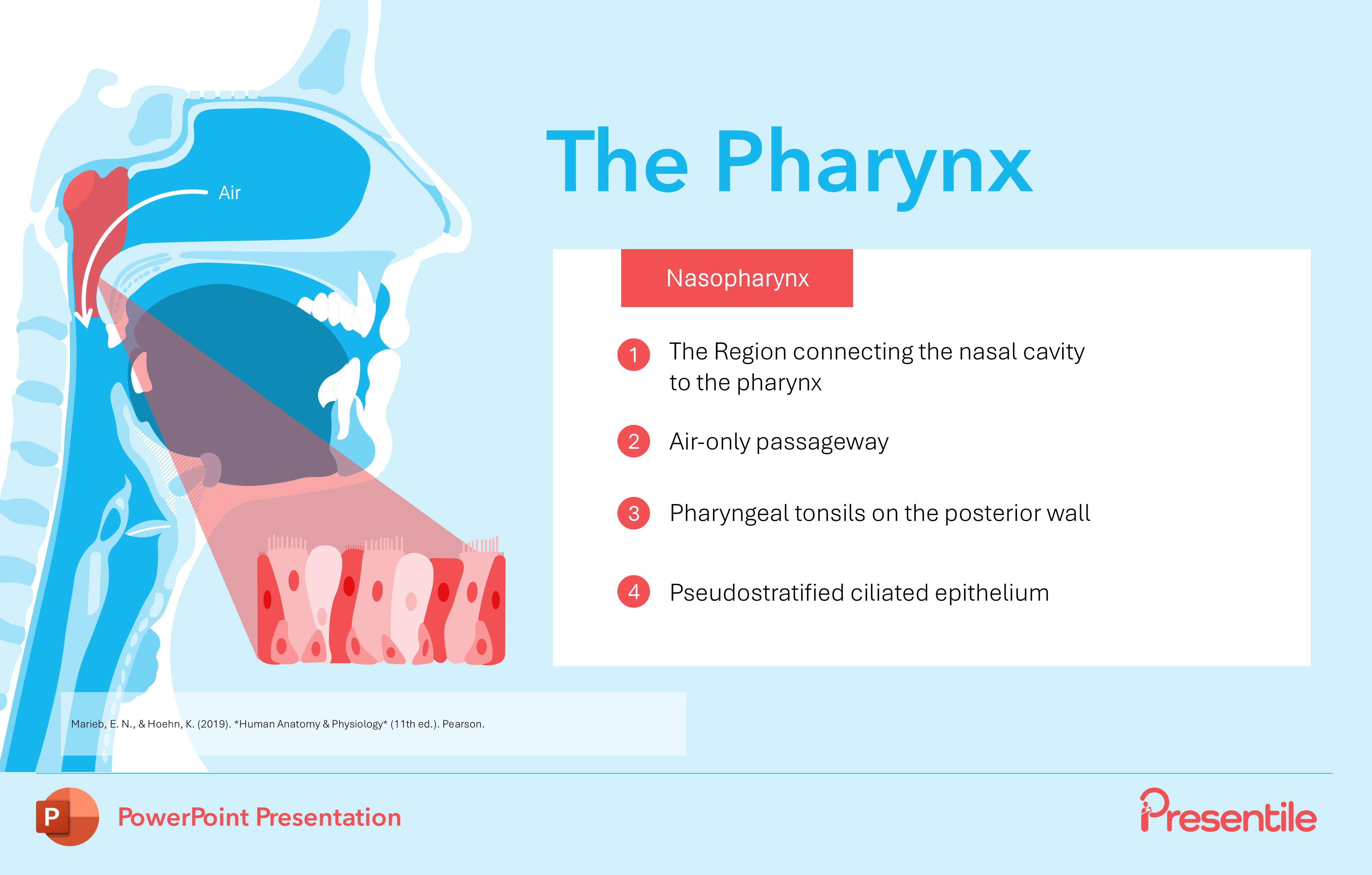
- This slide provides a detailed breakdown of the nasopharynx, the superior-most portion of the pharynx.
- It uses a numbered list to clearly outline its key characteristics, emphasizing that it serves exclusively as an air-only passageway.
- Furthermore, it delves into the microscopic anatomy, mentioning the presence of the pharyngeal tonsils and its lining of pseudostratified ciliated epithelium, giving the viewer a comprehensive understanding of this critical region.
Slide 23: The Oropharynx

- This slide focuses on the oropharynx, the second region of the pharynx.
- It provides a detailed overview of this critical area, highlighting its function as a shared passageway for both food and air.
- The slide also specifies its key anatomical features, including the presence of stratified squamous epithelium, and identifies the location of both the palatine and lingual tonsils, offering a comprehensive look at this section's structure and contents.
Slide 24: The Laryngopharynx
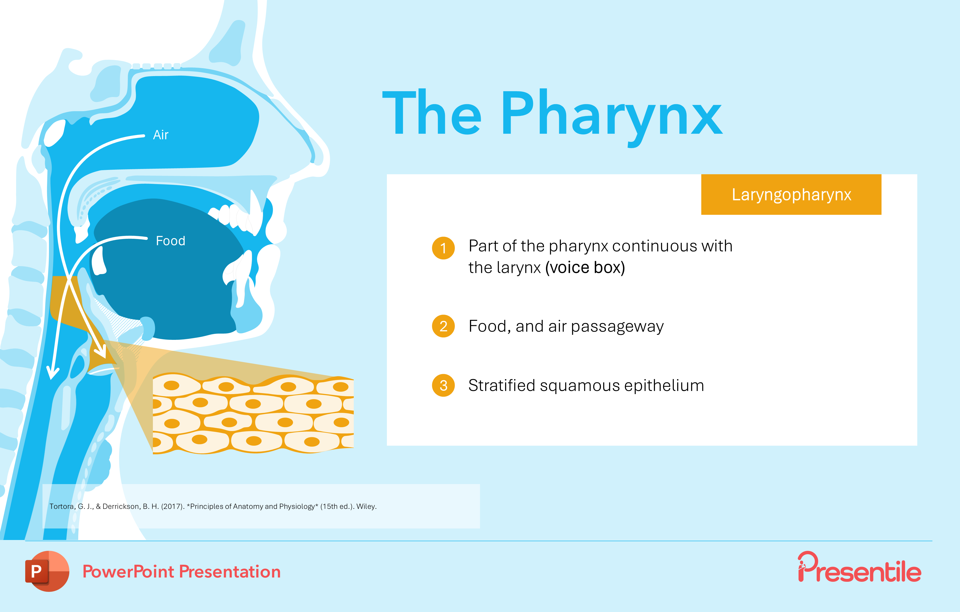
- This slide concludes the pharynx breakdown by detailing the laryngopharynx, the third and final region.
- It clearly outlines its anatomical position as the part of the pharynx continuous with the larynx, and reinforces its function as a shared passageway for both food and air.
- The slide also highlights the tissue lining this region, identifying it as stratified squamous epithelium, which solidifies the comprehensive and detailed nature of this anatomical presentation.
Slide 25: The Larynx

This slide introduces the larynx, a crucial structure commonly known as the voice box, marking a new section in the presentation. It provides an immediate overview of its primary function: connecting the pharynx to the trachea. The slide also highlights the key components it houses, including the vocal cords and the epiglottis, all supported by a clear, labeled anatomical diagram that identifies the major cartilages, offering a solid foundation for understanding this organ.
Slide 26: The Larynx cont.

This slide provides crucial anatomical context by detailing the exact location of the larynx within the neck. It uses a clear, numbered list to pinpoint its position relative to the cervical vertebrae (C3 to C6) and its connections to the hyoid bone superiorly and the trachea inferiorly. This information is fundamental to understanding the larynx's role as a passageway and its relationship to surrounding structures.
Slide 27: The Larynx Cartilages
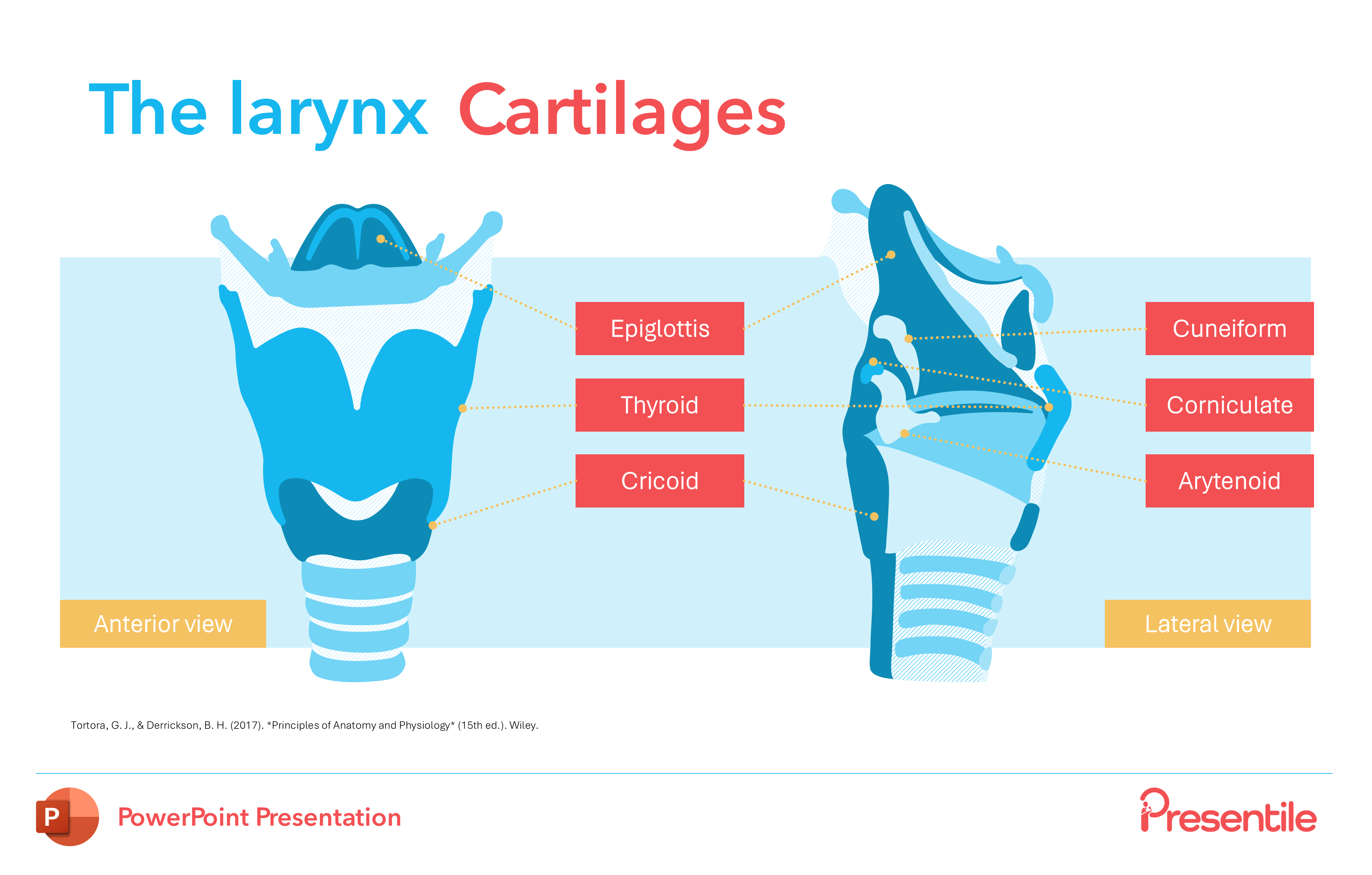
This slide provides a comprehensive anatomical breakdown of the cartilaginous framework of the larynx. It features both an anterior and a lateral view of the larynx, allowing for a complete three-dimensional understanding. The diagrams are clearly labeled to identify all major cartilages, including the epiglottis, thyroid, cricoid, cuneiform, corniculate, and arytenoid cartilages, making a complex anatomical structure easy to visualize and comprehend.
Slide 28: The Larynx Functions
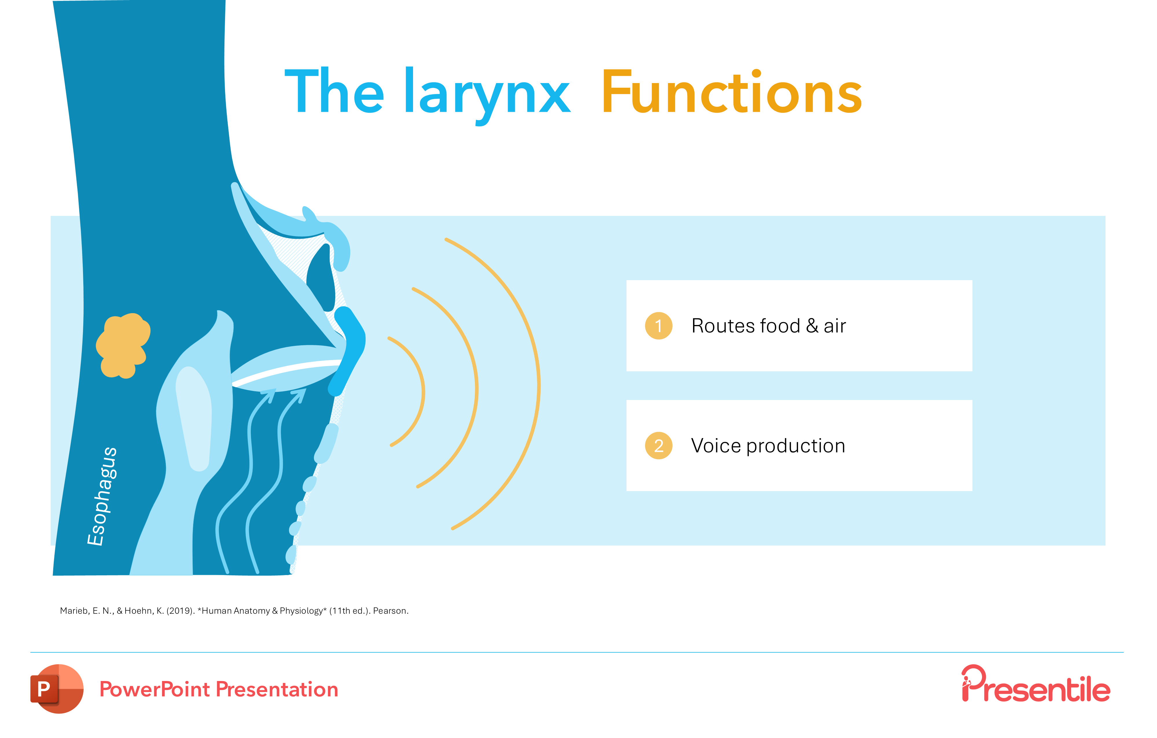
This slide succinctly details the two primary functions of the larynx. It uses numbered points and a clear, dynamic diagram to illustrate how the larynx acts as a critical passageway that routes food and air into their correct respective tracts. It also introduces the larynx's role in voice production, offering a concise yet comprehensive overview of the organ's essential physiological duties.
Slide 29: Vocal Cords
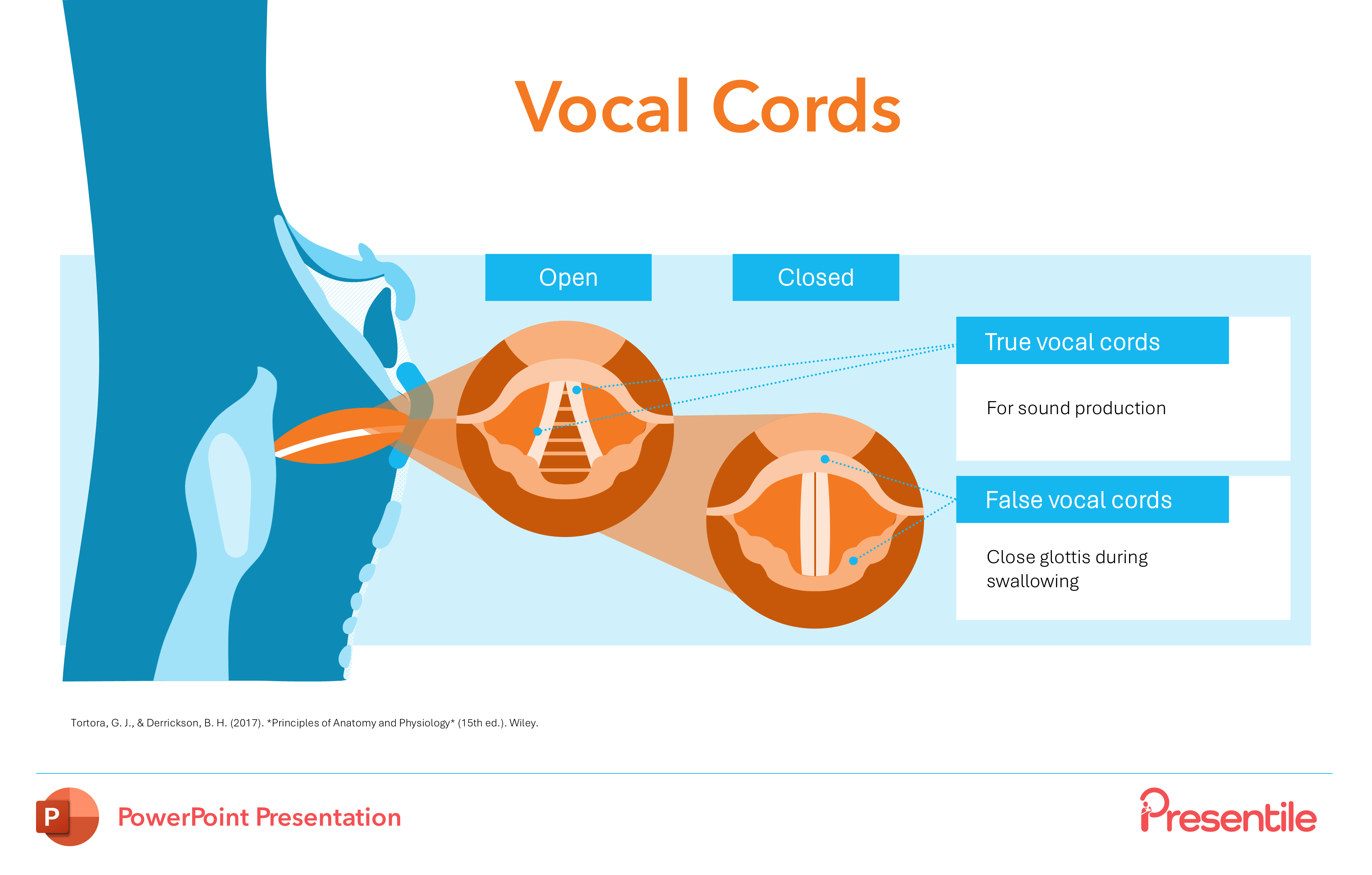
This slide provides a detailed look at the anatomy and function of the vocal cords, a core component of the larynx. It clearly differentiates between the true vocal cords, which are responsible for sound production, and the false vocal cords, which function to close the glottis during swallowing. The slide's side-by-side diagrams visually illustrate the "open" and "closed" states of the vocal cords, offering a clear and dynamic understanding of how they perform their distinct roles in both speech and protecting the airway.
Slide 30: The Lower Respiratory Tract

This slide serves as a clear transition to a new major section of the presentation, introducing the Lower Respiratory Tract. The visual on the slide immediately orients the viewer, showing a high-level overview of the lungs, bronchi, and bronchioles. This powerful introductory slide sets the stage for a detailed anatomical and functional exploration of the respiratory system below the larynx.
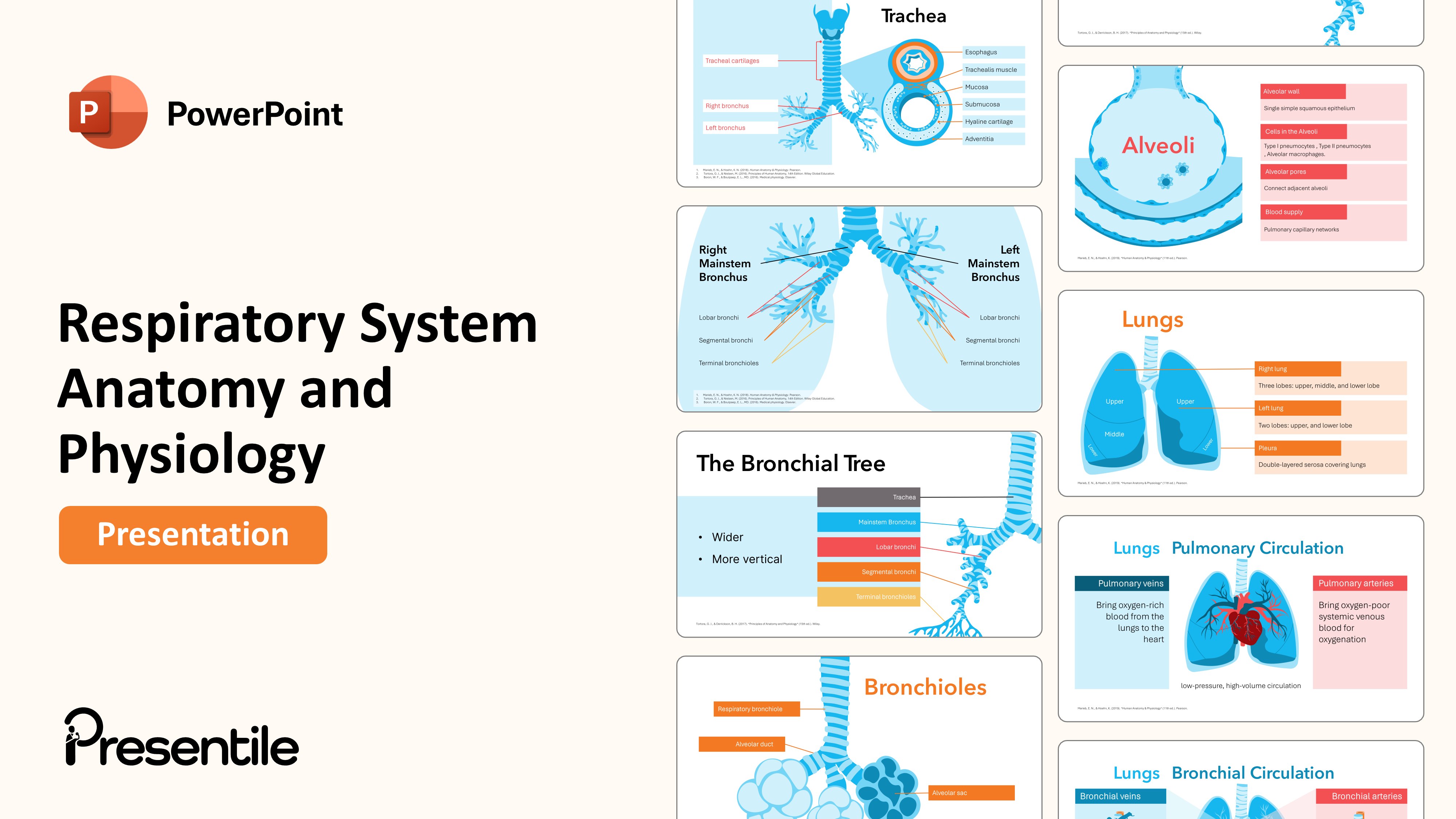
Slide 31: The Trachea
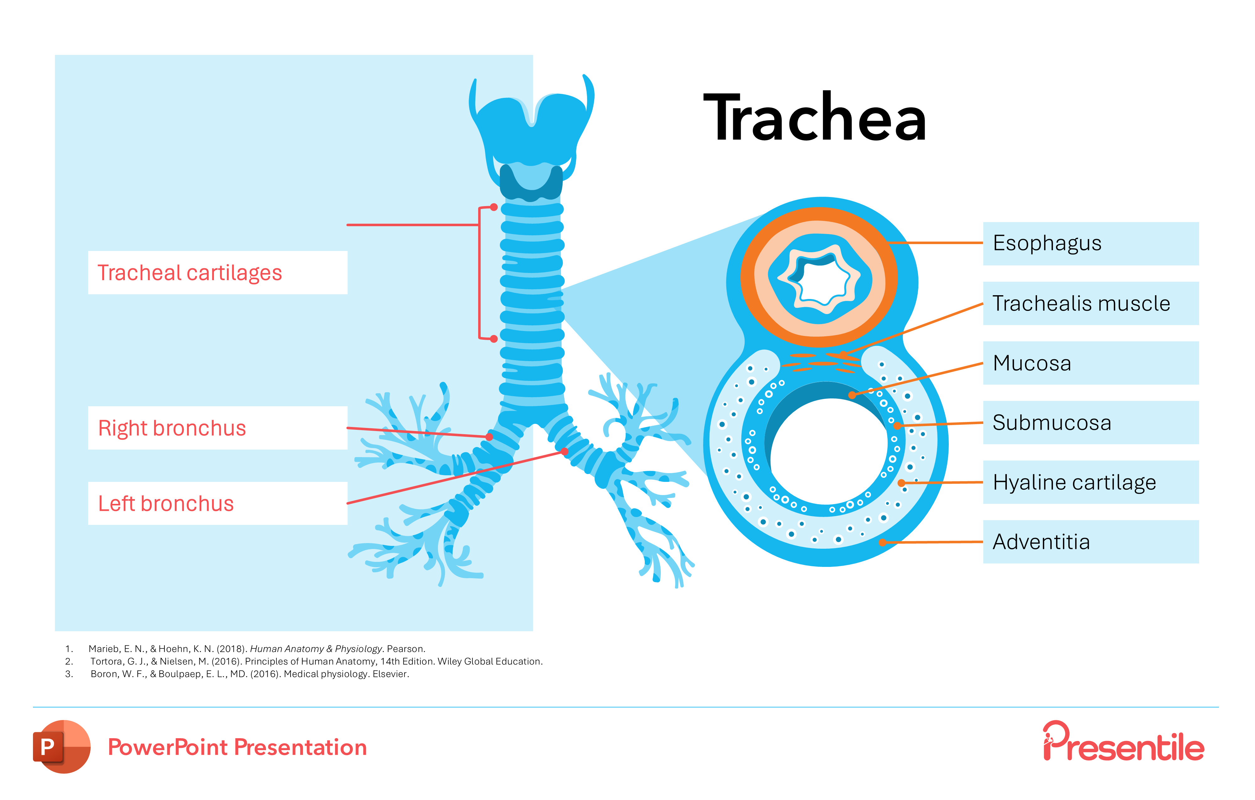
This slide provides a comprehensive view of the trachea, detailing both its overall structure and microscopic anatomy. It features an anatomical diagram showing the trachea's division into the right and left bronchus, along with a detailed cross-section of the tracheal wall. This cross-section clearly labels the layers, including the mucosa, submucosa, and hyaline cartilage, offering an in-depth look at this vital airway.
Slide 32: The Bronchial Tree
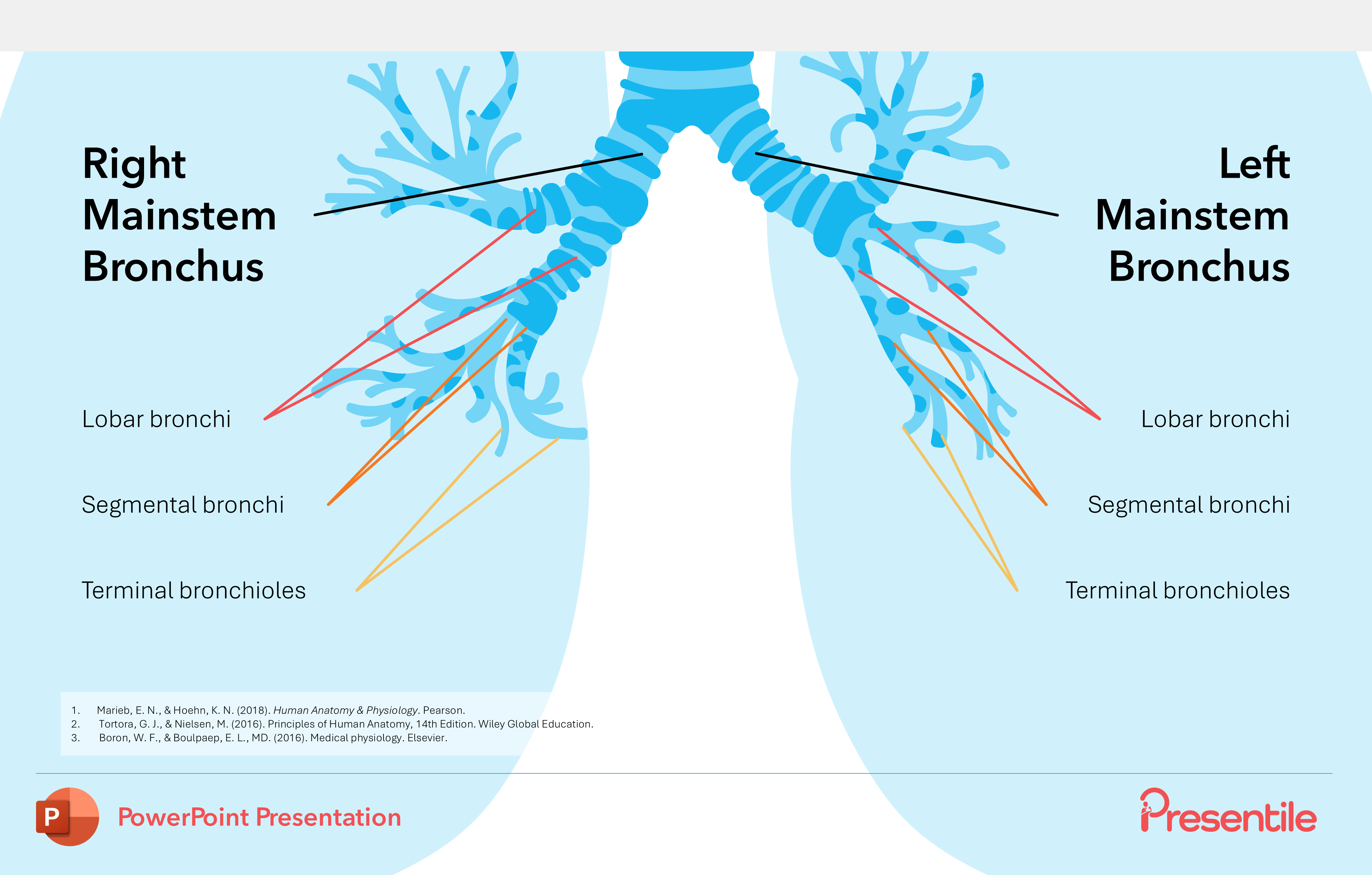
This slide provides a detailed and organized overview of the bronchial tree, the complex network of airways within the lungs. The clear, anatomical diagram visually breaks down the hierarchy of the airways, labeling the Right and Left Mainstem Bronchus, the Lobar bronchi, the Segmental bronchi, and the Terminal bronchioles. This slide is essential for understanding how air is efficiently distributed throughout the lungs.
Slide 33: The Mainstem Bronchus
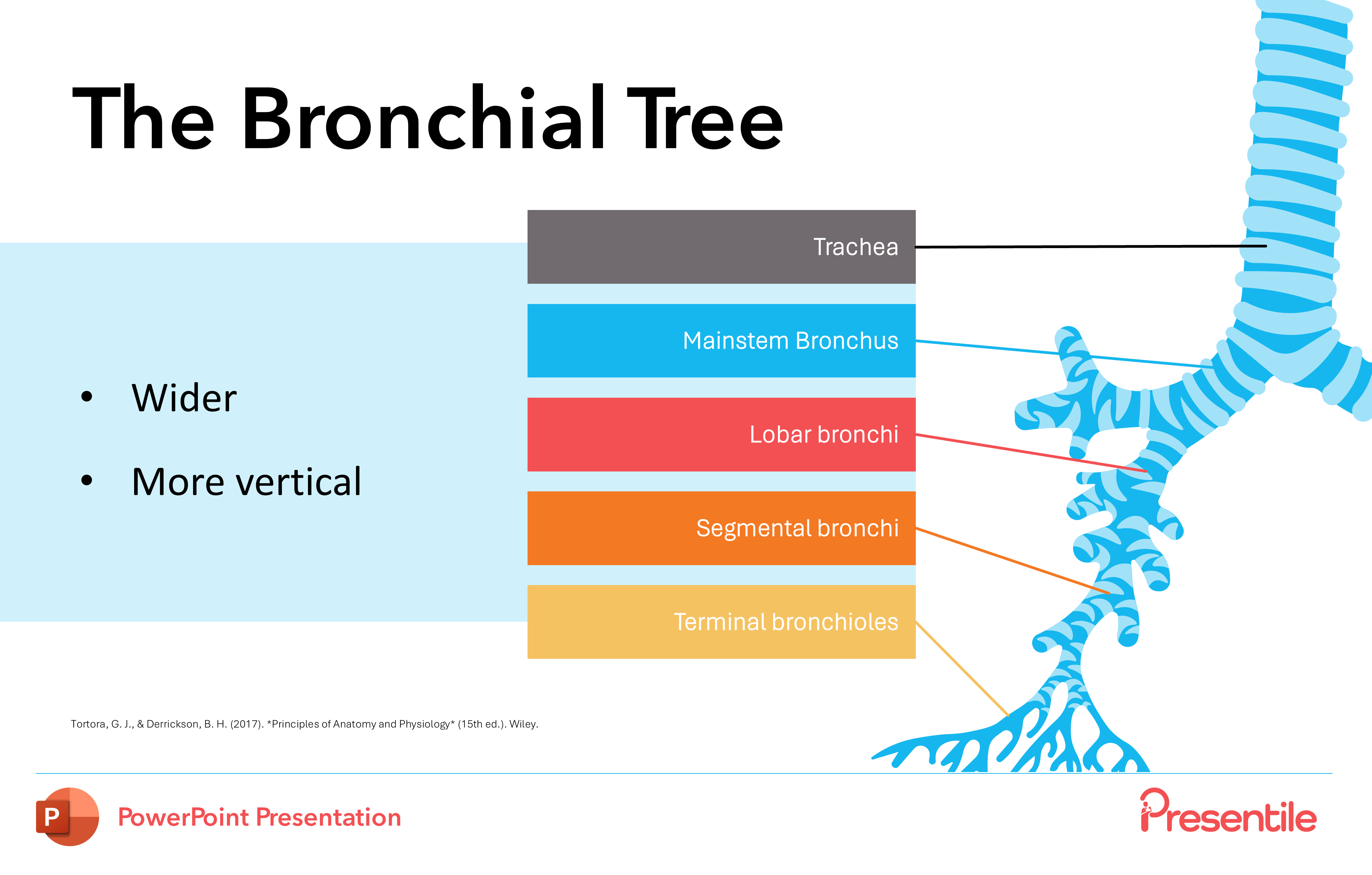
This slide provides a more detailed look at the anatomy of the mainstem bronchus. It clearly highlights the key characteristic that the right mainstem bronchus is wider and more vertical than the left, a crucial anatomical detail for clinical applications. This focus on specific details demonstrates the presentation's depth, moving beyond a simple overview to provide essential, practical knowledge.
Slide 34: The Upper Bronchial Tree
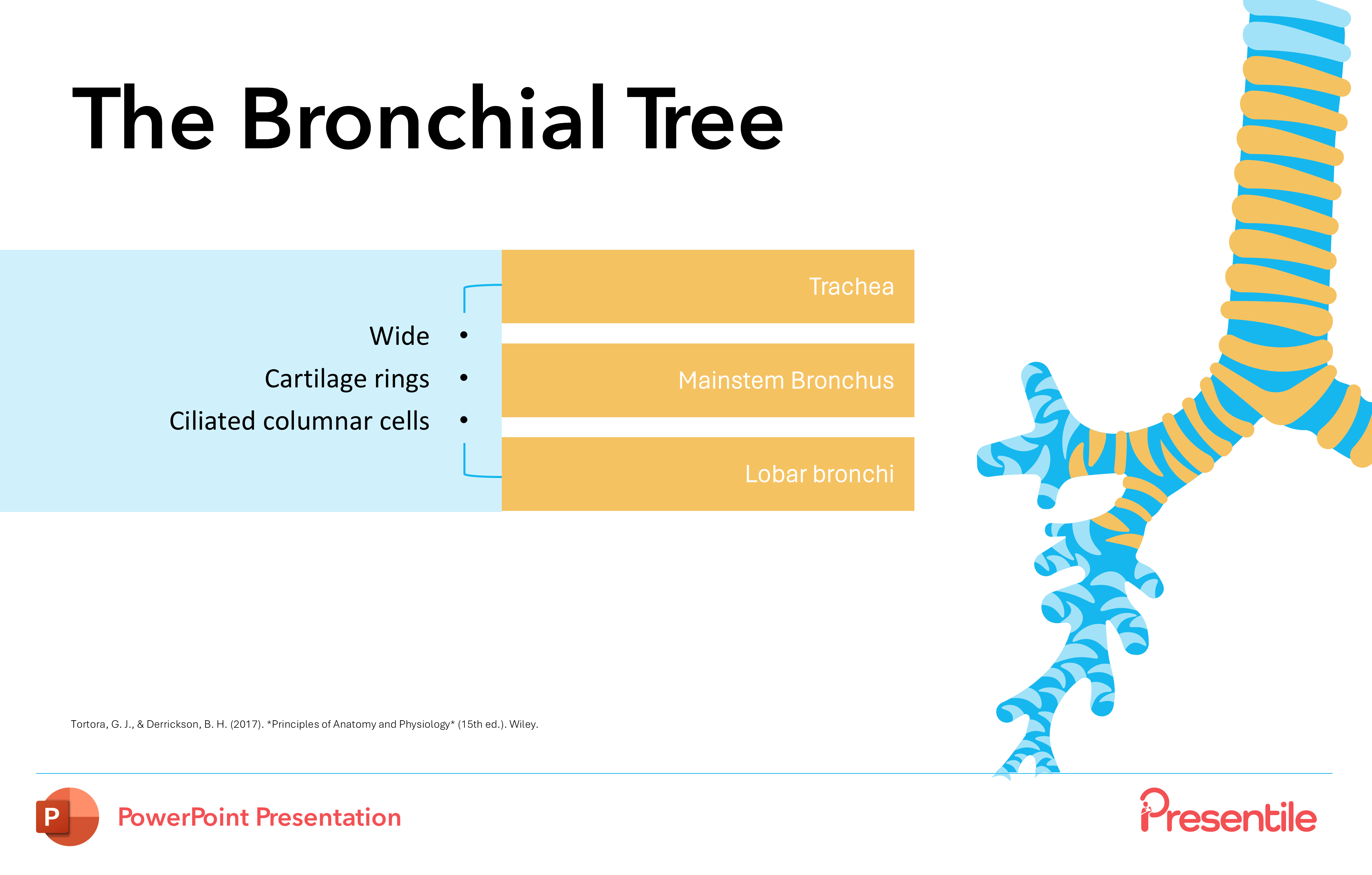
This slide provides a detailed look at the structural components of the upper bronchial tree. It highlights key features of the mainstem bronchus, including its wide diameter, the presence of cartilage rings for structural support, and the lining of ciliated columnar cells, which are essential for clearing debris from the airways. This focus on both macroscopic and microscopic detail provides a comprehensive understanding of the anatomy.
Slide 35: The Bronchioles
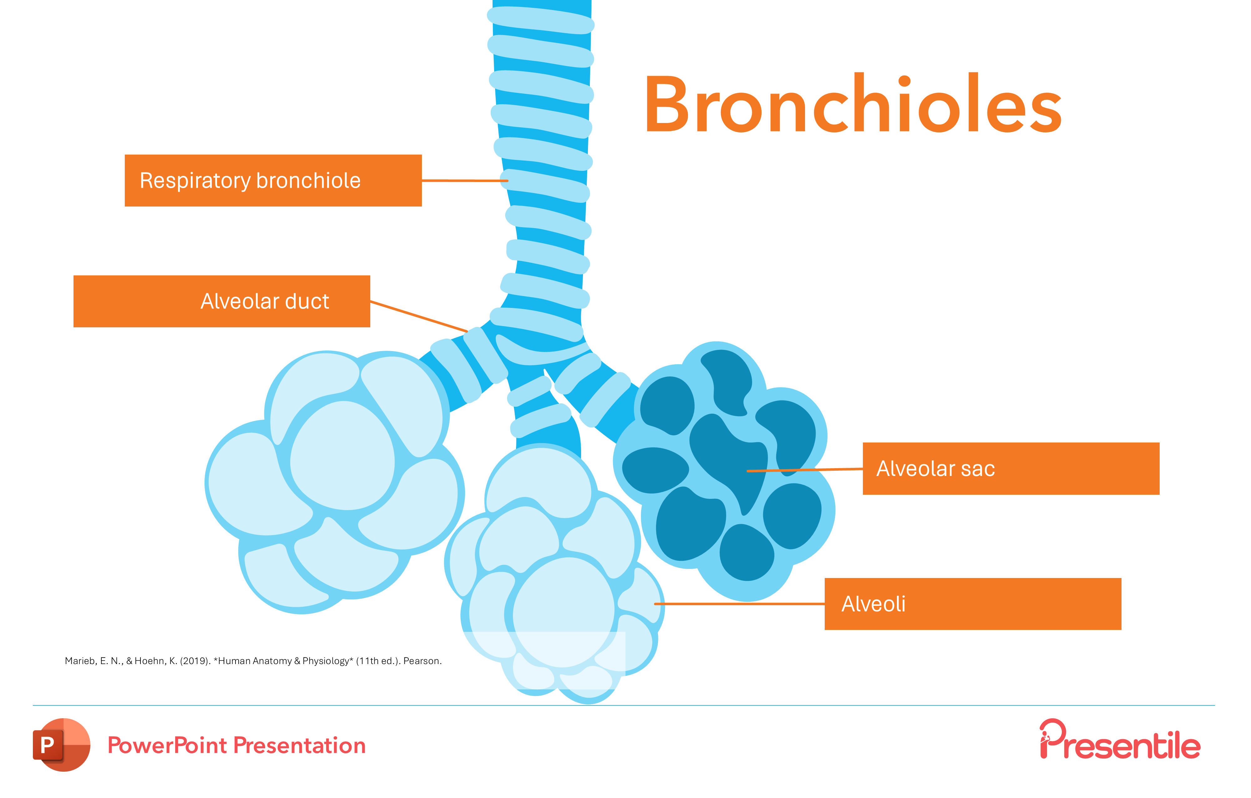
This slide marks a crucial transition in the presentation, moving into the final and most critical structures of the respiratory tree. It provides a detailed diagram that illustrates the progression from the respiratory bronchiole to the alveolar ducts, sacs, and alveoli. This section is essential for understanding where the vital process of gas exchange takes place.
Slide 36: The Alveoli
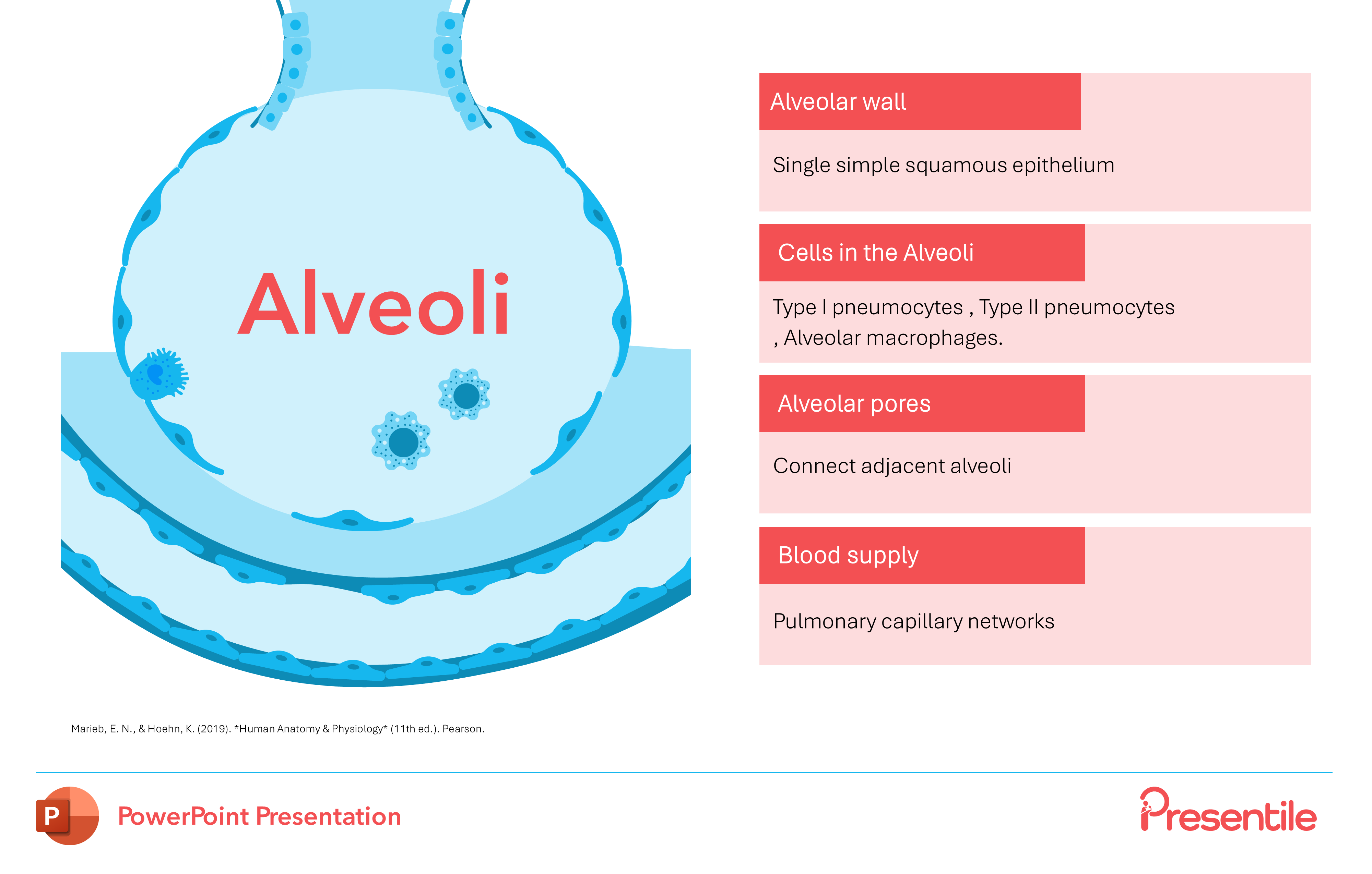
This slide provides a micro-anatomical deep dive into the alveoli, the primary functional units of the lungs. It details the cellular composition of the alveolar wall with a focus on its single simple squamous epithelium, and identifies the different cell types crucial for function: Type I and Type II pneumocytes and alveolar macrophages. The slide also explains the purpose of alveolar pores and highlights the vital blood supply from the pulmonary capillary networks, offering a complete picture of gas exchange at the cellular level.
Slide 37: The Lungs
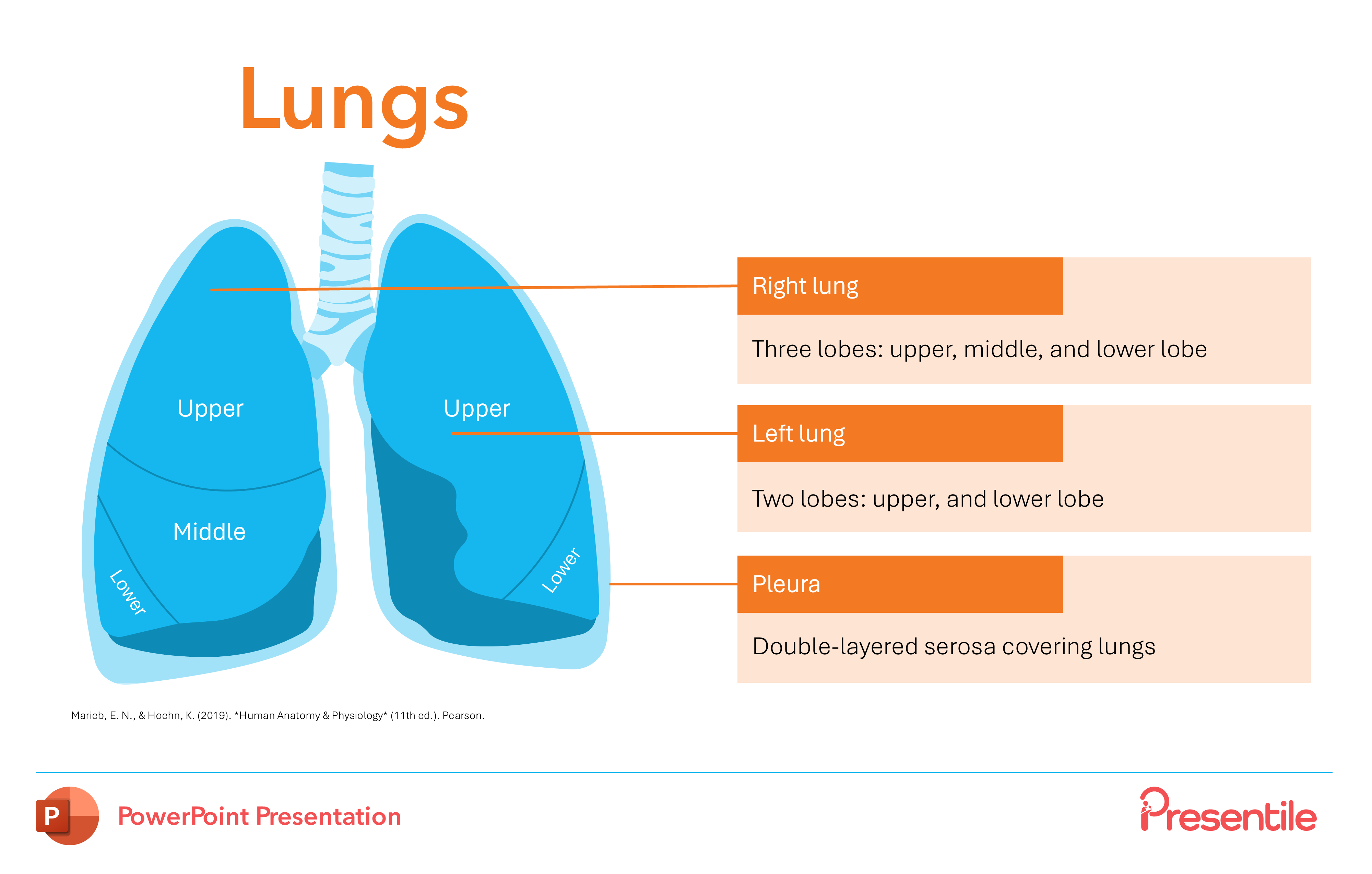
This slide provides an essential overview of the lungs' gross anatomy. It uses a clear, color-coded diagram to visually distinguish the right lung with its three lobes and the left lung with its two lobes. The slide also concisely defines the pleura as the double-layered serosa covering the lungs, offering a complete picture of the external structure and protective coverings.
Slide 38: The Lungs Pulmonary Circulation

This slide provides an essential overview of the pulmonary circulation, illustrating how the lungs are integrated with the cardiovascular system. It clearly distinguishes the functions of the pulmonary arteries that carry oxygen-poor blood to the lungs from the pulmonary veins that return oxygen-rich blood to the heart. This segment is vital for understanding the complete process of gas exchange and reinforces the interconnectedness of the body's systems.
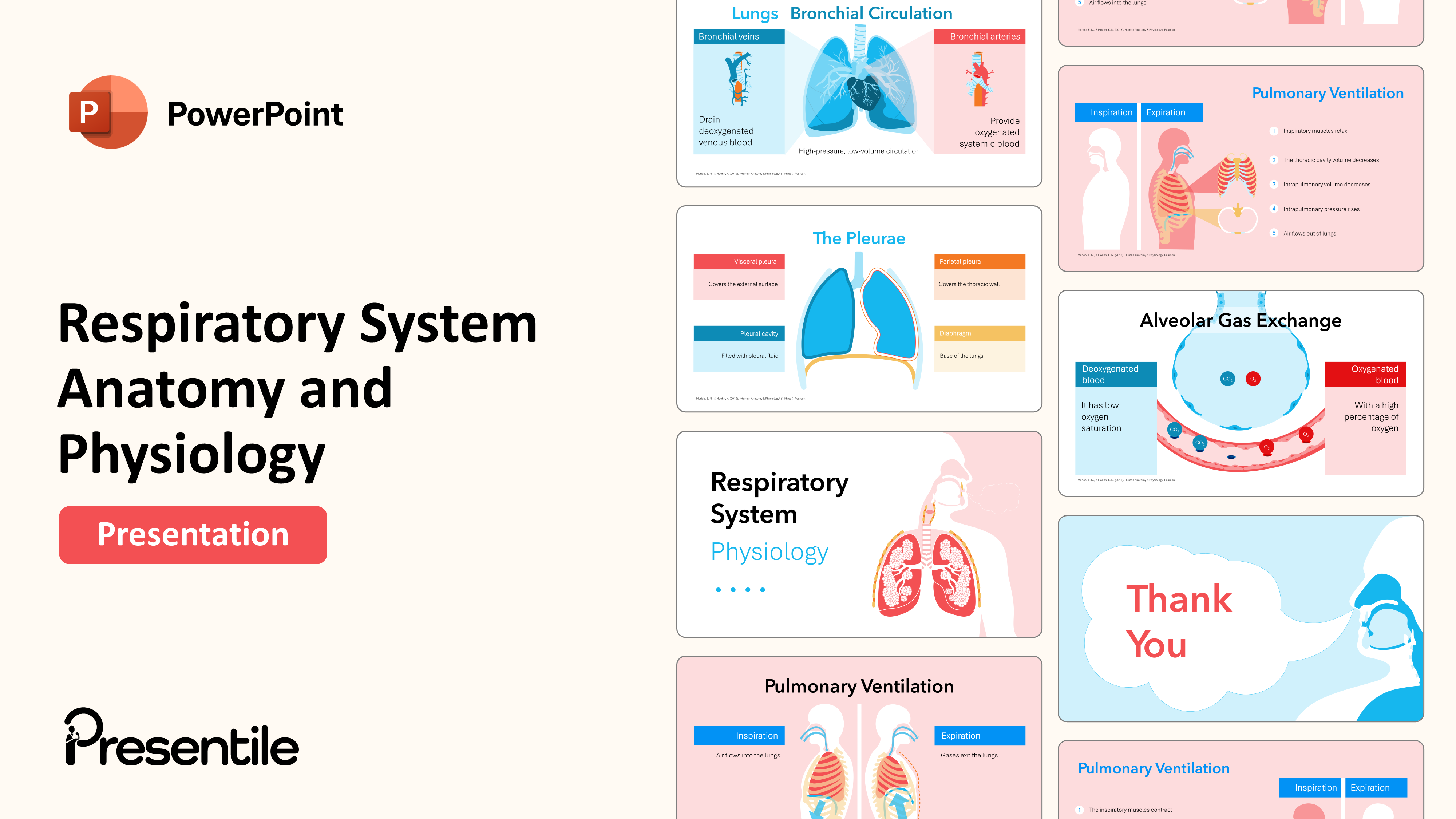
Slide 39: Lungs Bronchial Circulation
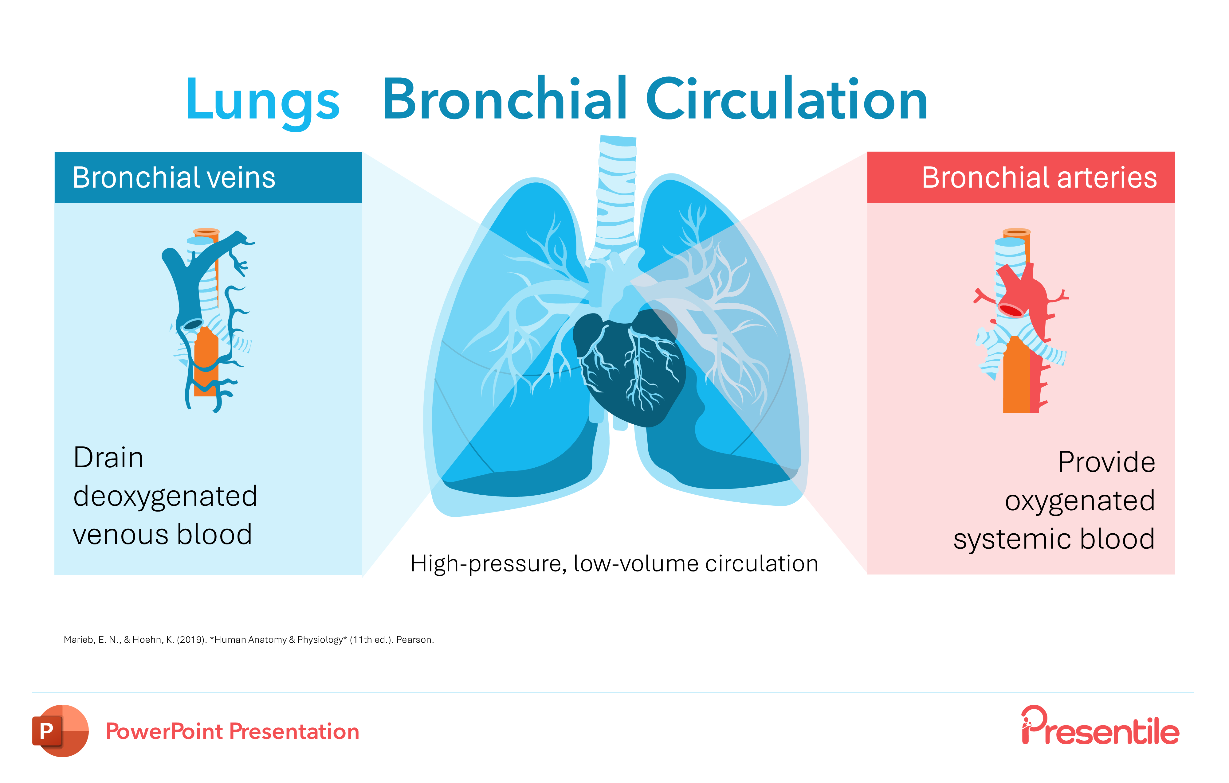
This slide distinguishes the bronchial circulation from the pulmonary system, providing critical detail on how the lung tissue itself is supplied with blood. It clearly explains that bronchial arteries provide oxygenated blood directly to the lungs' structures, while bronchial veins drain the deoxygenated blood. This distinction highlights the presentation's comprehensive coverage of both the functional and nutritional blood supplies of the respiratory system.
Slide 40: The Pleurae

This slide provides a focused look at the pleural membranes and their role in lung function. It clearly differentiates between the visceral pleura, which covers the external surface of the lungs, and the parietal pleura, which lines the thoracic wall. The slide also explains the purpose of the pleural cavity and the fluid it contains, offering a complete picture of the protective layers and the mechanics of breathing.
Slide 41: Respiratory System Physiology

This slide marks a significant transition, serving as an introductory slide to a new section focused on respiratory system physiology. It signals a shift from the anatomical structures to a detailed exploration of how the system functions. This slide sets the stage for a deep dive into the mechanics of breathing, gas exchange, and the regulation of respiration.
Slide 42: Pulmonary Ventilation

This slide initiates the physiology section by explaining the fundamental mechanics of pulmonary ventilation, or breathing. It uses a clear, side-by-side diagram to illustrate the processes of both inspiration and expiration, showing how the movement of the diaphragm and thoracic wall facilitates the flow of air into and out of the lungs. This visual explanation simplifies a complex physiological process.
Slide 43: Pulmonary Ventilation (Inspiration)
.PNG)
This slide provides a step-by-step breakdown of the process of inspiration. It uses a clear, numbered list alongside an illustrative diagram to explain the sequence of events, from the contraction of the inspiratory muscles and the subsequent increase in thoracic cavity volume to the drop in intrapulmonary pressure that ultimately draws air into the lungs. This detailed, sequential approach clarifies the physical principles behind breathing.
Slide 44: Pulmonary Ventilation ( Expiration)
.PNG)
This slide provides a detailed, step-by-step explanation of the process of expiration. It uses a numbered list to clarify the sequence of events, beginning with the relaxation of the inspiratory muscles and the subsequent decrease in thoracic cavity volume. This leads to a rise in intrapulmonary pressure that ultimately forces air out of the lungs, effectively completing the discussion on the mechanics of breathing.
Slide 45: Alveolar Gas Exchange
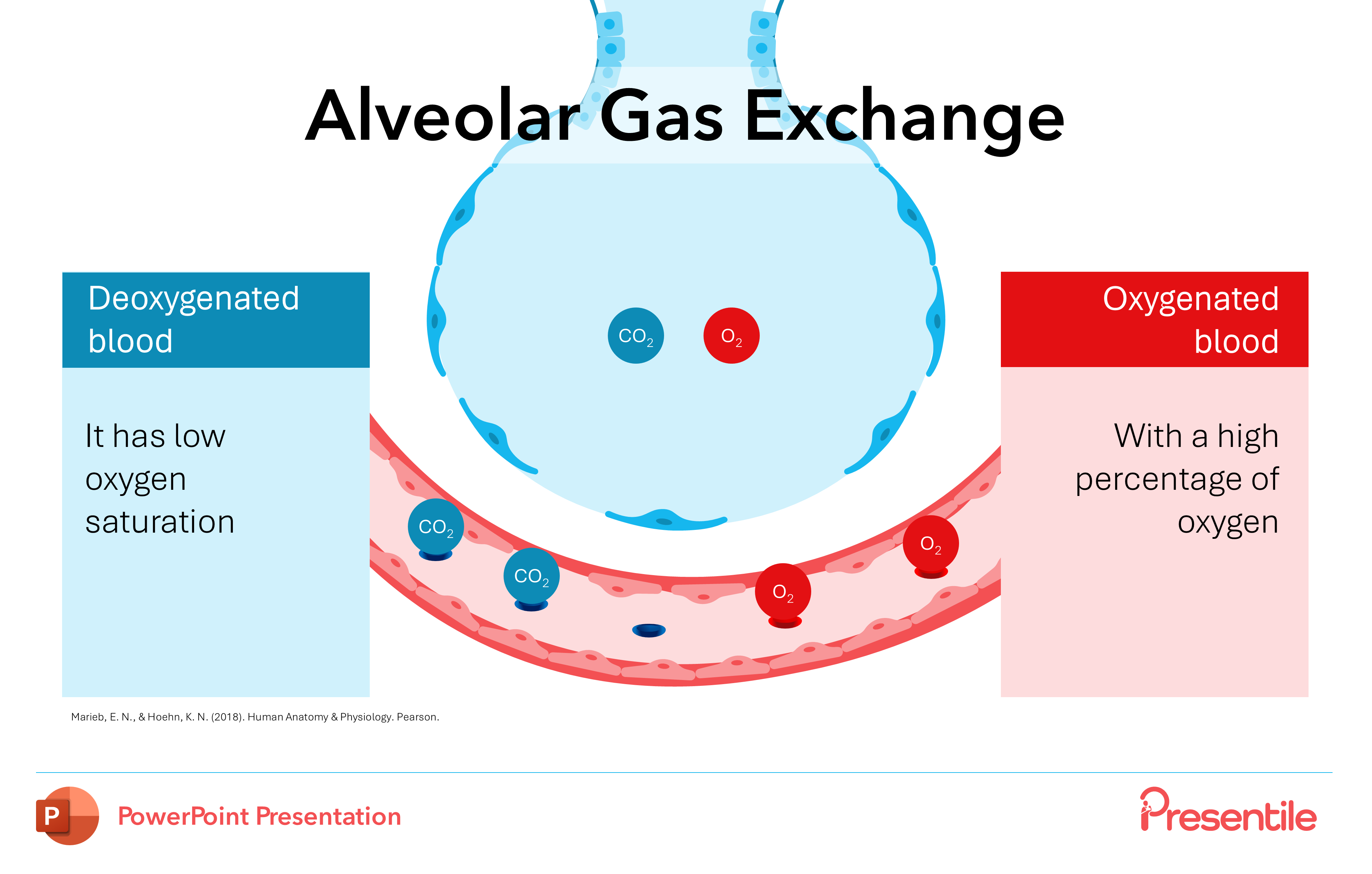
This slide provides a clear visual explanation of alveolar gas exchange, the vital process occurring in the lungs. It uses a detailed diagram to illustrate how deoxygenated blood, with its low oxygen saturation, releases carbon dioxide and takes up oxygen as it passes by the alveoli, becoming oxygenated blood. This dynamic slide effectively demonstrates the key physiological event that the entire respiratory system is built to perform.
Slide 46: Thank You Slide

This presentation offers a complete and visually rich exploration of the respiratory system, from the initial anatomy of the nasal cavity and pharynx to the complex physiology of the lungs. Its comprehensive approach, which includes detailed breakdowns of structures, step-by-step explanations of processes like pulmonary ventilation and gas exchange, and high-quality diagrams, makes it an invaluable educational tool for a wide range of audiences. The presentation effectively transitions between macro- and micro-level anatomy and physiology, providing a holistic understanding of how the respiratory system functions.
Features of
Respiratory System Physiology & Anatomy PowerPoint Presentation
- Fully editable in PowerPoint
- All graphics are in vector format
- Medically Referenced information and data
Respiratory System Physiology & Anatomy PowerPoint Presentation
Preview Presentation Slides count:
 Compatible with:Microsoft PowerPoint
 File type:PPTX
 Dimensions:16:9
- Non-animated PowerPoint
- Animated PowerPoint File
- Animated PowerPoint with Voice Over
- PDF Documents with presentation script


