Medical Infographics
Respiratory
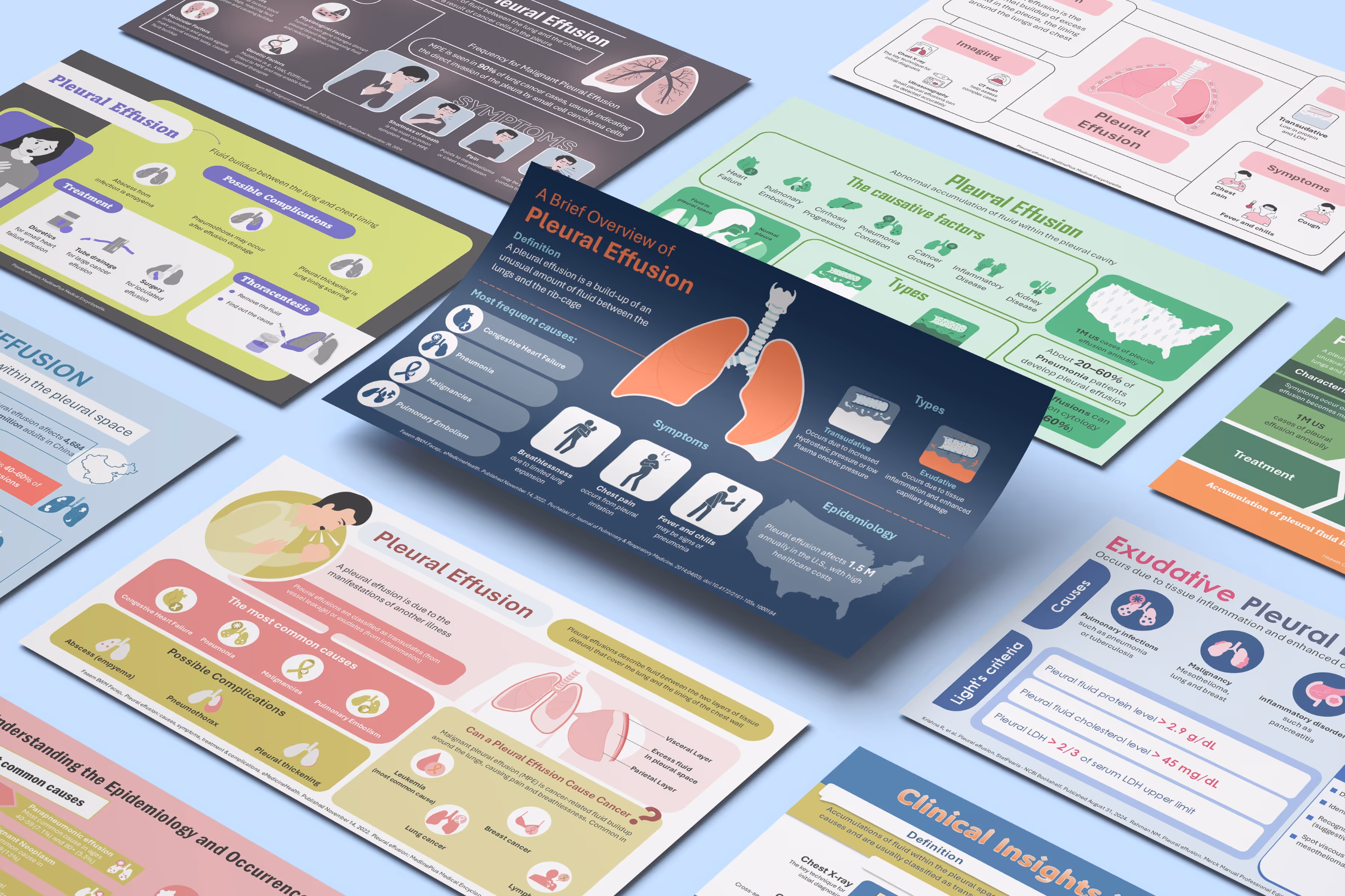
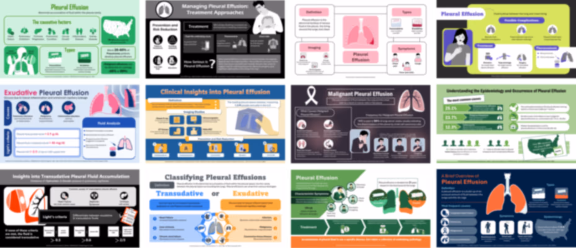
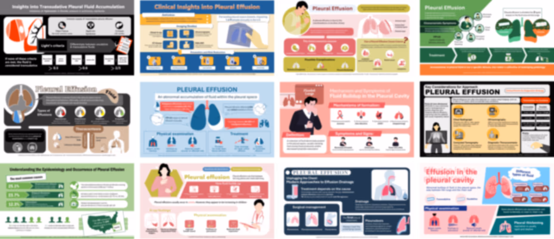
Pleural Effusion PowerPoint Infographics
No items found.


Trusted By Life Scinece Teams:


Fully editable in PowerPoint with customizable colors, shapes, and layouts
For easy and friendly drag and drop
Fully animated slides
To help simplify and communicate complex medical concepts
All graphics in vector format
For crisp visuals at any size
Medically referenced information and data
Built by medical professionals and researchers who perfectly understand the science behind the slides
Life sciences–focused design
Tailored for pharma, biotech, and healthcare organizations
Price:
$ 49.00 USD
For pharmaceutical marketing teams and medical affairs teams

- who need to explain pleural effusion clearly without building visuals from scratch, these ready-made infographics help turn complex clinical information—such as causes, classification, diagnosis, and treatment—into simple, visual content that is easy to present and understand, while being fully editable and designed to fit directly into any PowerPoint slide and match your existing presentation colors automatically.
For training managers, healthcare organizations, hospitals, and clinical training programs

- looking to make sessions more engaging, these infographics help simplify complex respiratory topics, making it easier for learners to follow key concepts like fluid accumulation, classification, and management, without requiring additional design work.
For professors, academic lecturers, and medical education programs

- who want to support teaching with clear visuals, these infographics break down pleural effusion into structured visual sections—covering mechanisms, symptoms, imaging, and treatment—helping students grasp the topic faster while allowing flexibility to integrate into lectures.
For healthcare professionals including pulmonologists, internal medicine physicians, and clinical educators
- who need to explain concepts quickly, these visuals support clearer communication during clinical discussions, patient education, or presentations, helping reduce explanation time while improving understanding.
For medical communication agencies, consultants, and healthcare educators
- who need reusable visual content, these infographic slides can be easily adapted, reused, and integrated into presentations, educational materials, or digital content without redesigning from scratch.
No items found.