.webp)
No items found.
Content of
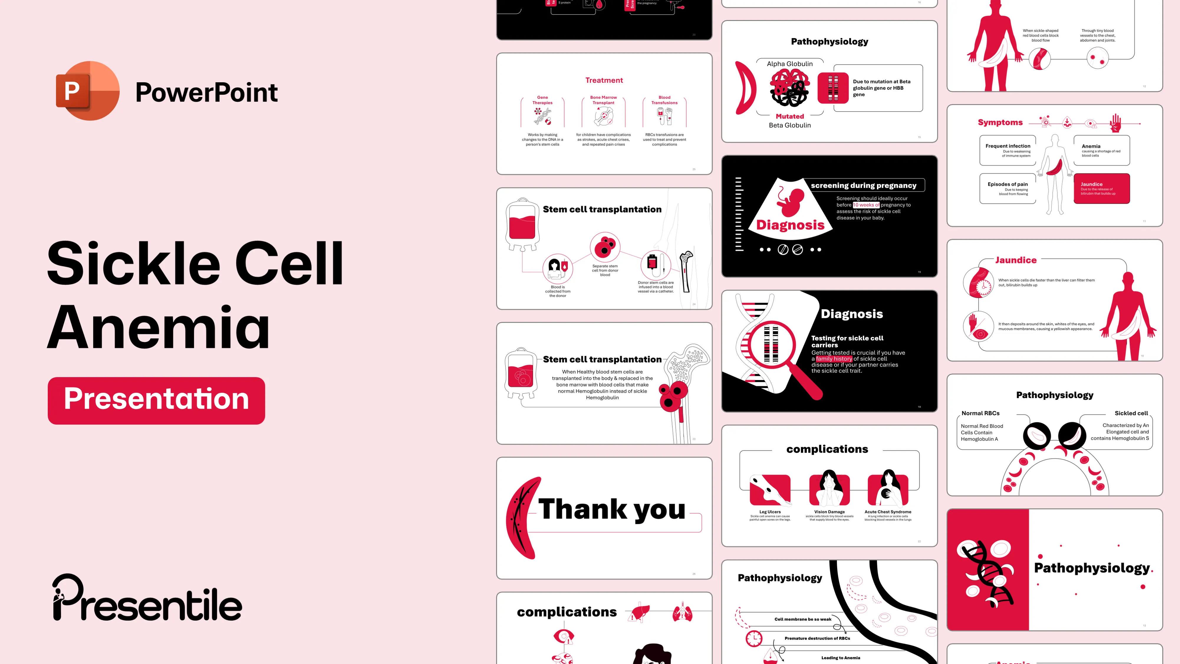
Sickle Cell Anemia Presentation Monochrome
Slide 1: Introduction to Sickle Cell Anemia

- This title slide introduces the comprehensive presentation on Sickle Cell Anemia.
- The design features a stylized depiction of a sickle-shaped red blood cell, instantly communicating the core topic.
- This visual element, combined with the clear, bold title, immediately focuses the audience's attention and sets the stage for a detailed medical overview of the disease.
- It provides a strong, professional starting point for a presentation that promises to be both informative and easy to follow.
Slide 2: The Pathophysiology of Sickle Cell Anemia
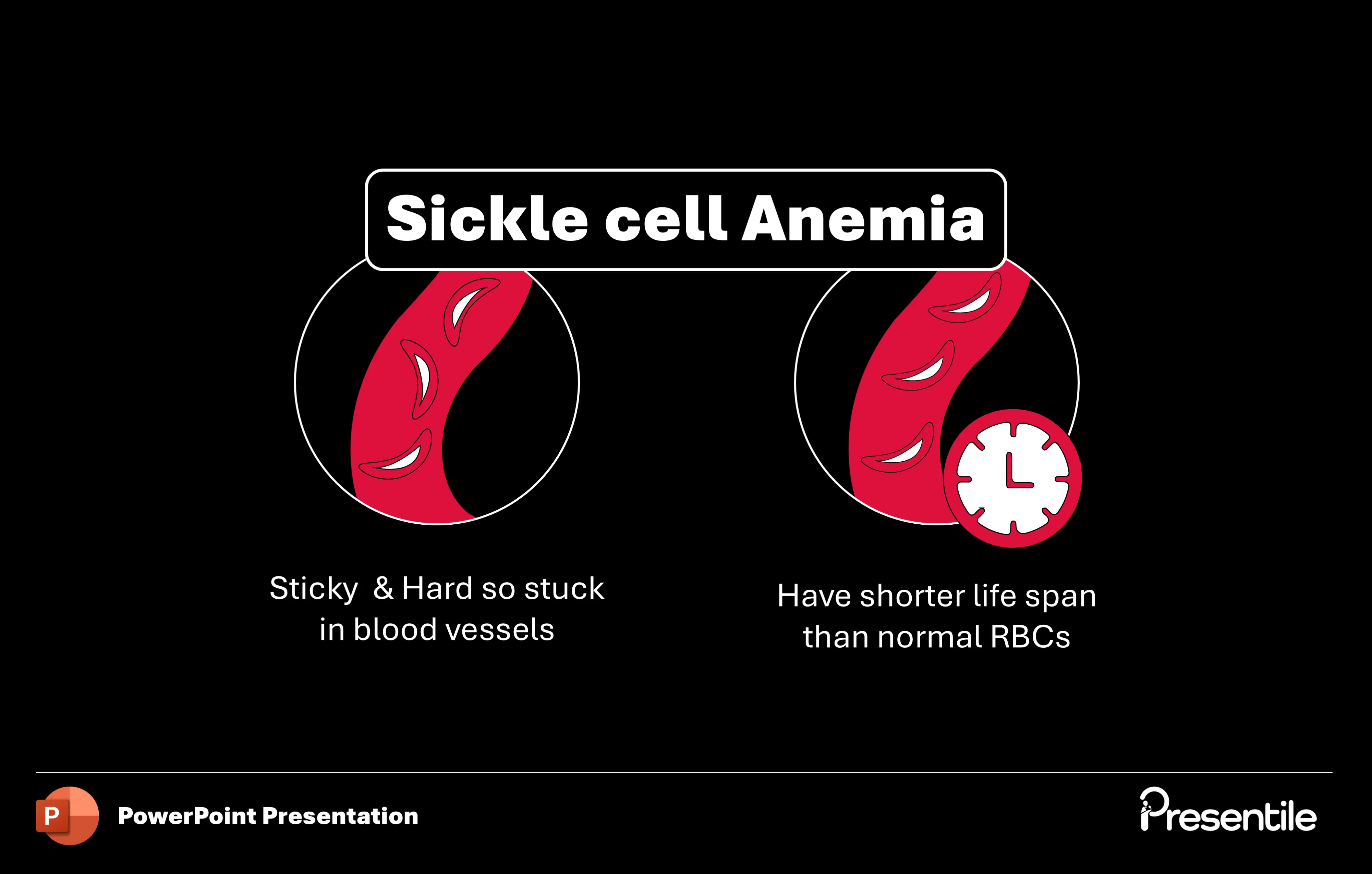
- This slide breaks down the core characteristics of sickle cell anemia.
- It uses two clear visual aids to illustrate the key problems.
- The first shows the rigid, sticky nature of sickle-shaped red blood cells, explaining how they get stuck in blood vessels.
- The second visual, featuring a clock icon, highlights the shortened lifespan of these abnormal cells compared to normal red blood cells. Together, these graphics and concise text simplify the complex pathophysiology of the disease, making it easy for the audience to grasp the fundamental mechanics of blood flow disruption and anemia.
Slide 3: Key Facts About Sickle Cell Anemia

- This slide provides a high-level overview of essential facts about Sickle Cell Anemia.
- It uses a bold, red background and a clean layout to present four key points, each with a distinct icon.
- These include the long-term nature of the disease, the potential for dangerous complications, its genetic link (Family History), and the necessity of lab testing for diagnosis.
- The slide is designed for quick comprehension, summarizing critical information in an easy-to-digest, visually engaging format.
Slide 4: Epidemiology of Sickle Cell Anemia
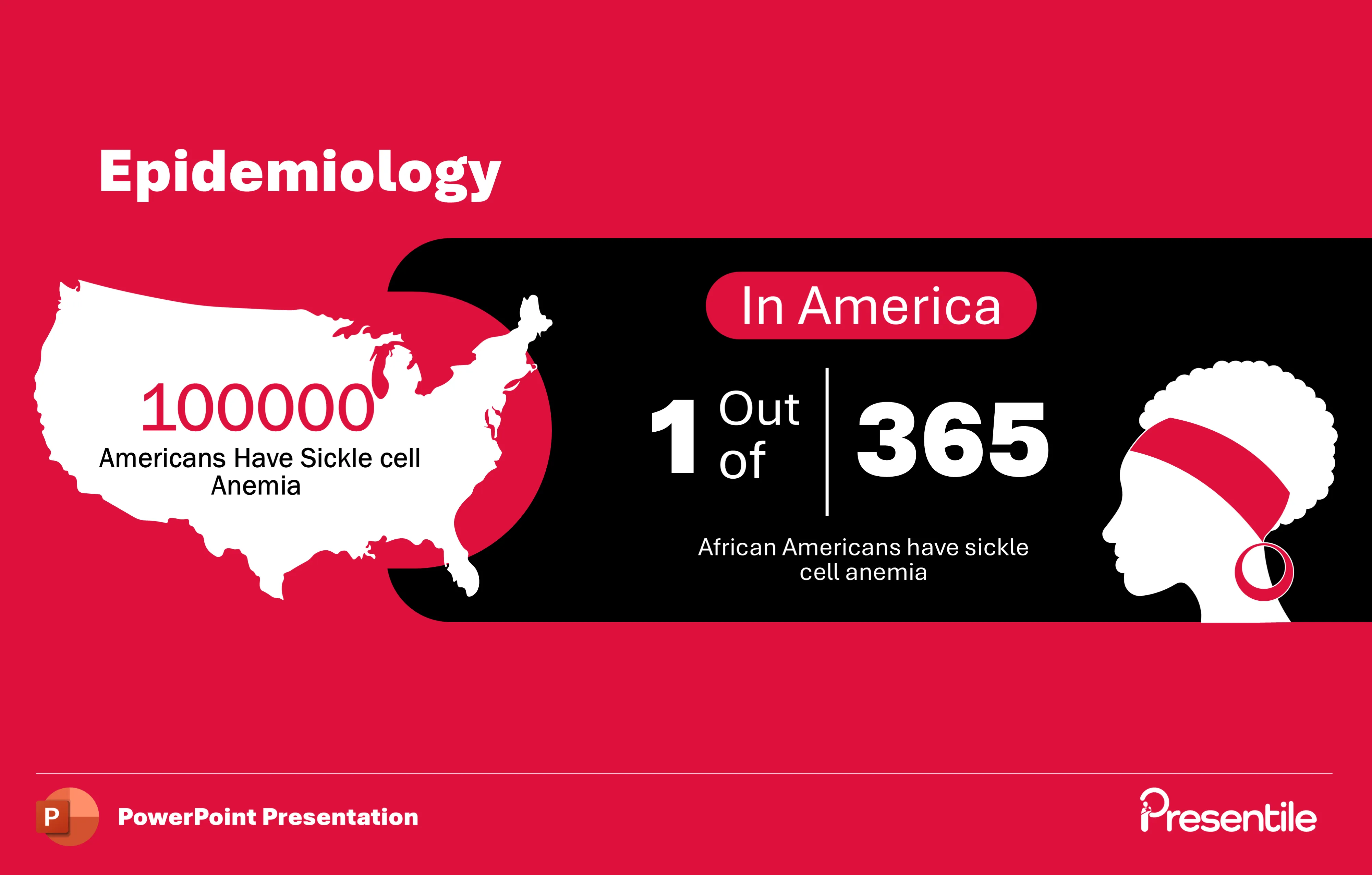
- This slide presents key epidemiological data on Sickle Cell Anemia in the United States.
- It uses a bold, red-and-black color scheme and impactful visuals to highlight the statistics.
- The first statistic, placed over a map of the United States, notes that 100,000 Americans have the disease.
- The second, more specific statistic, displayed prominently with a stylized silhouette, reveals that 1 in 365 African Americans are affected.
- This slide effectively communicates the prevalence and disproportionate impact of the disease, providing a crucial context for understanding its public health significance.
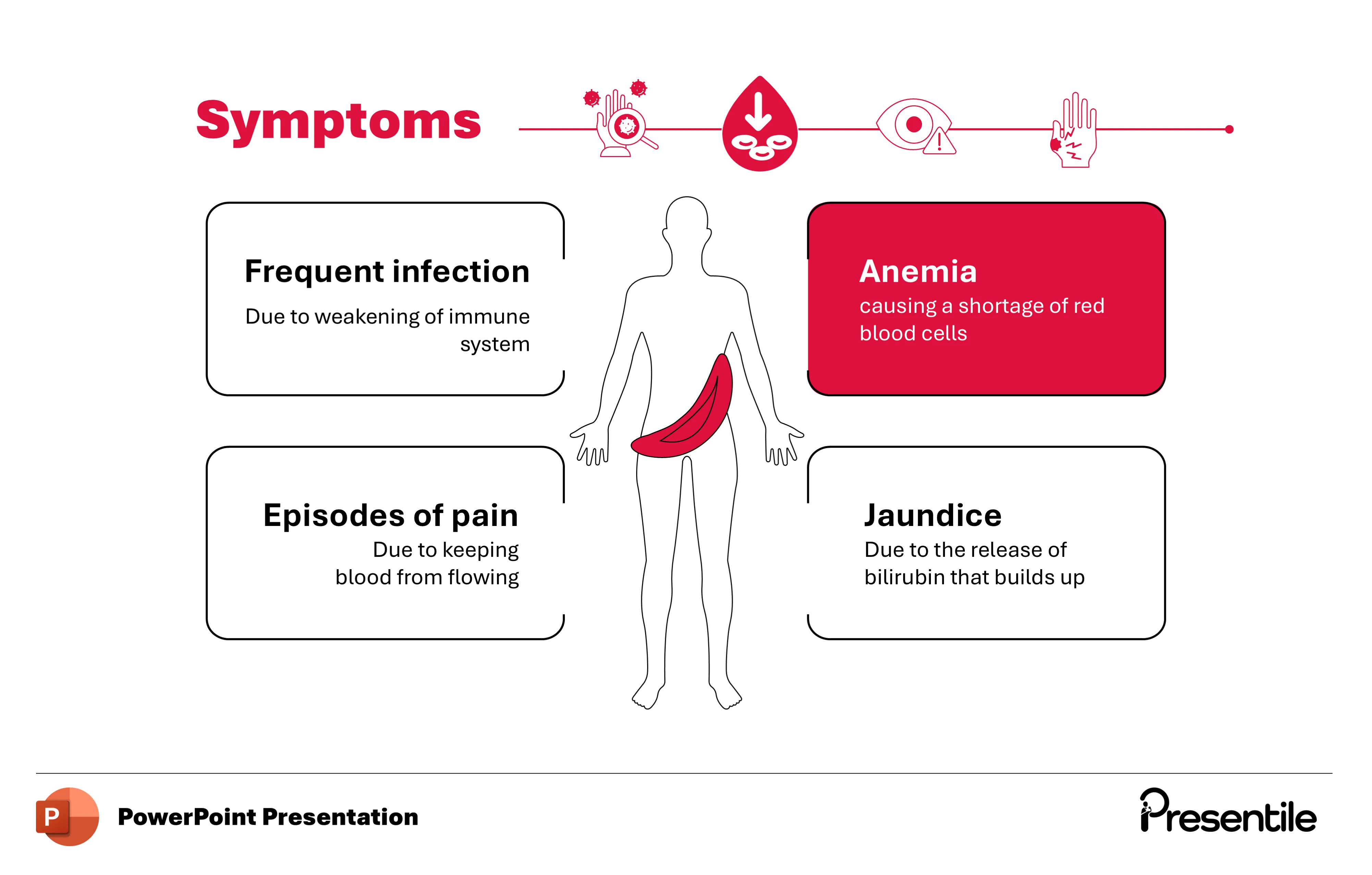
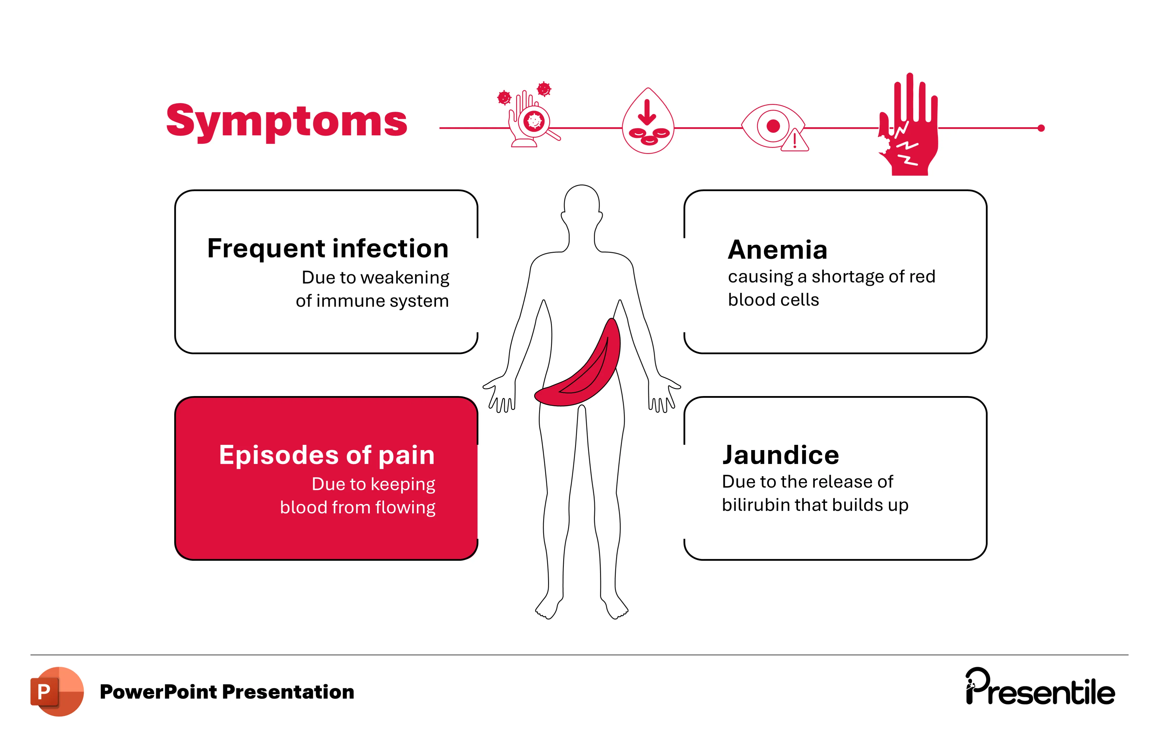
Slide 5: Key Symptoms of Sickle Cell Anemia
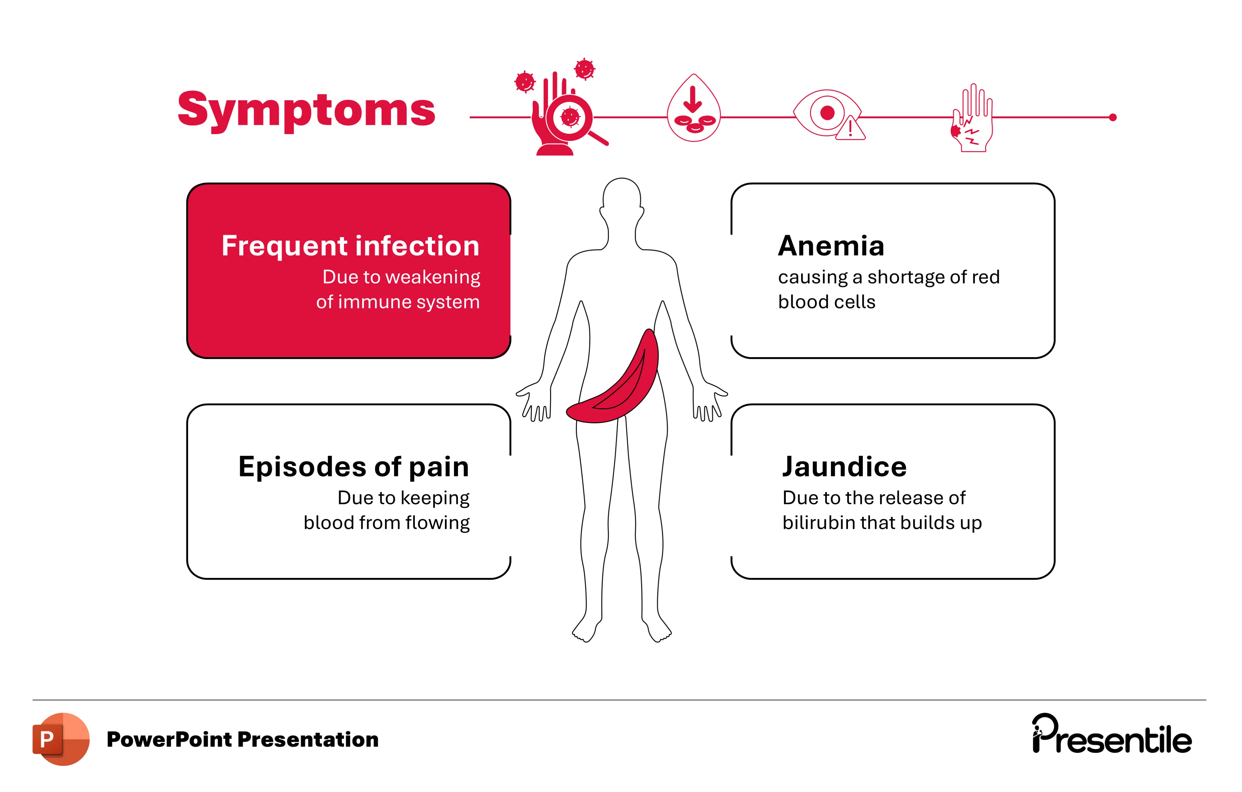
- This slide details the most common symptoms of Sickle Cell Anemia. A central human silhouette acts as an anchor for four distinct symptom boxes.
- The symptoms covered are Frequent infection, Anemia, Episodes of pain, and Jaundice, each with a brief explanation of its cause.
- The clean, connected design effectively links the symptoms to the affected body systems, making it easy for the audience to understand the multi-faceted clinical presentation of the disease.
- A row of icons at the top visually summarizes the different symptom categories, reinforcing the information presented.
Slide 6: Understanding Frequent Infections in Sickle Cell Anemia
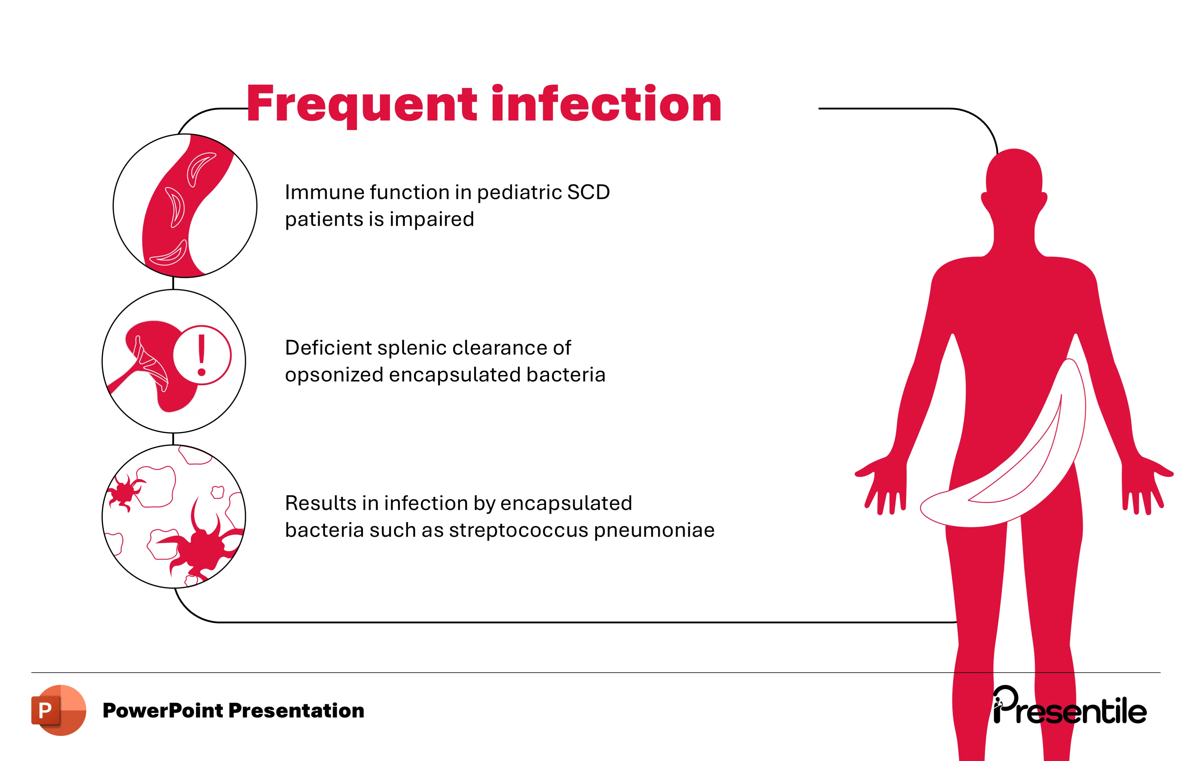
- This slide provides a detailed explanation of why patients with Sickle Cell Anemia, particularly children, experience frequent infections.
- The design uses a flowing, connected visual to illustrate the chain of events.
- It begins with the impaired immune function in pediatric patients and moves to the key mechanism: deficient splenic clearance of opsonized encapsulated bacteria.
- The final point clarifies that this deficiency leads to infections caused by specific bacteria, such as Streptococcus pneumoniae.
- This slide simplifies a complex immunological concept, offering a clear and concise breakdown of the pathophysiology behind a major complication of the disease.
Slide 7: Unpacking Anemia in Sickle Cell Disease
- This slide focuses on the symptom of Anemia, a central feature of Sickle Cell Disease.
- It uses a bold, red highlight to draw attention to this specific symptom box.
- The slide reinforces the concept that anemia is caused by the shortage of red blood cells that results from the disease.
- This concise explanation helps the audience understand that the core issue isn't just the sickle shape, but the body's inability to maintain a sufficient count of healthy, oxygen-carrying cells.
Slide 8: The Mechanism of Anemia in Sickle Cell Disease
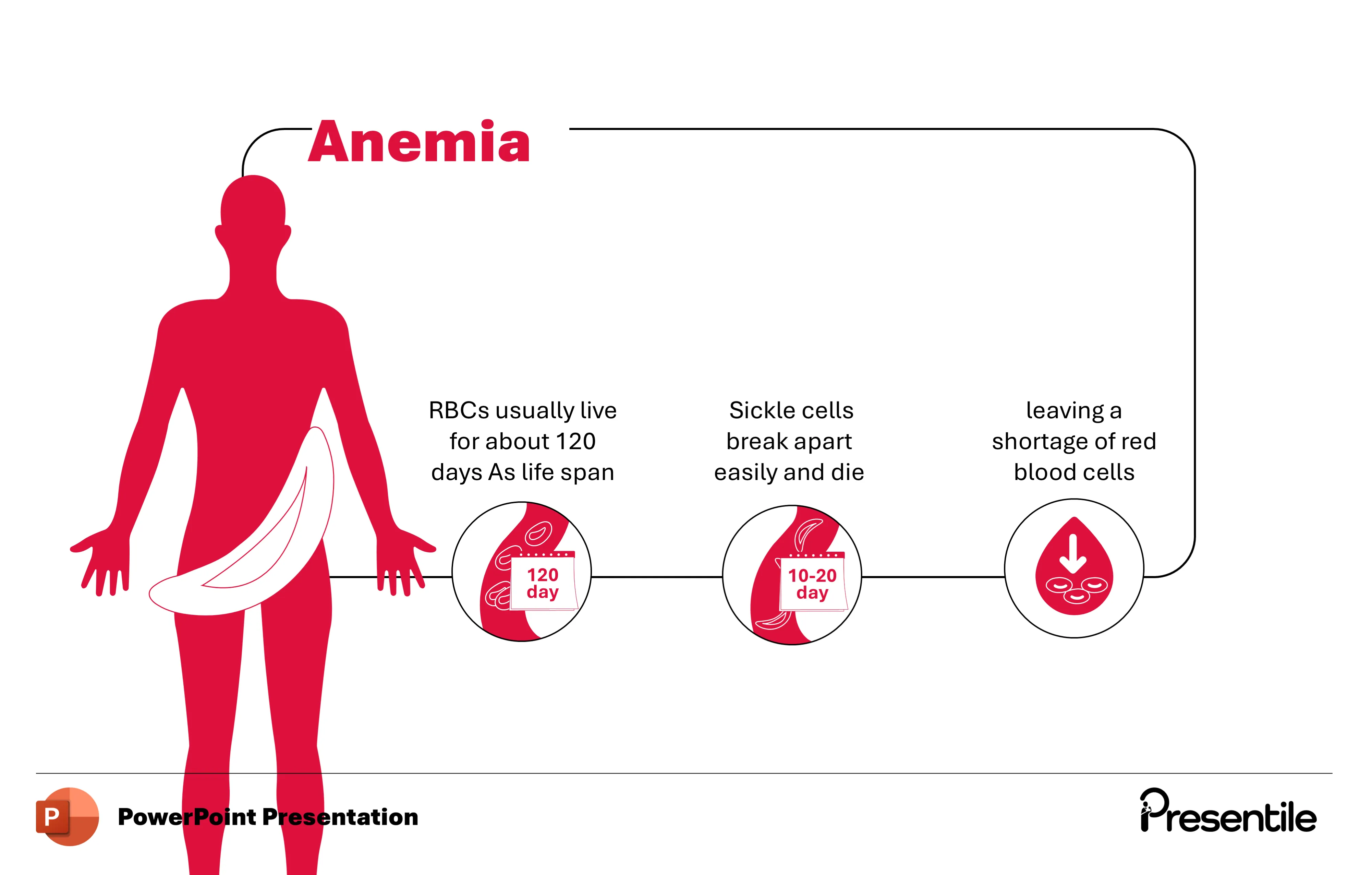
- This slide provides a detailed, step-by-step explanation of how anemia develops in Sickle Cell Anemia.
- It uses a horizontal flow to compare the lifespan of healthy red blood cells (RBCs) to that of sickle cells.
- The first visual shows a normal RBC lifespan of approximately 120 days.
- The second visual contrasts this by showing that sickle cells break apart easily and die much faster, living only 10 to 20 days.
- The final image depicts a downward-pointing arrow within a red blood cell, symbolizing the resulting shortage of red blood cells that causes anemia.
- This slide clearly illustrates the root cause of the patient's anemic state, reinforcing the concepts from previous slides with a clear, chronological breakdown.
Slide 9: Highlighting Jaundice in Sickle Cell Anemia
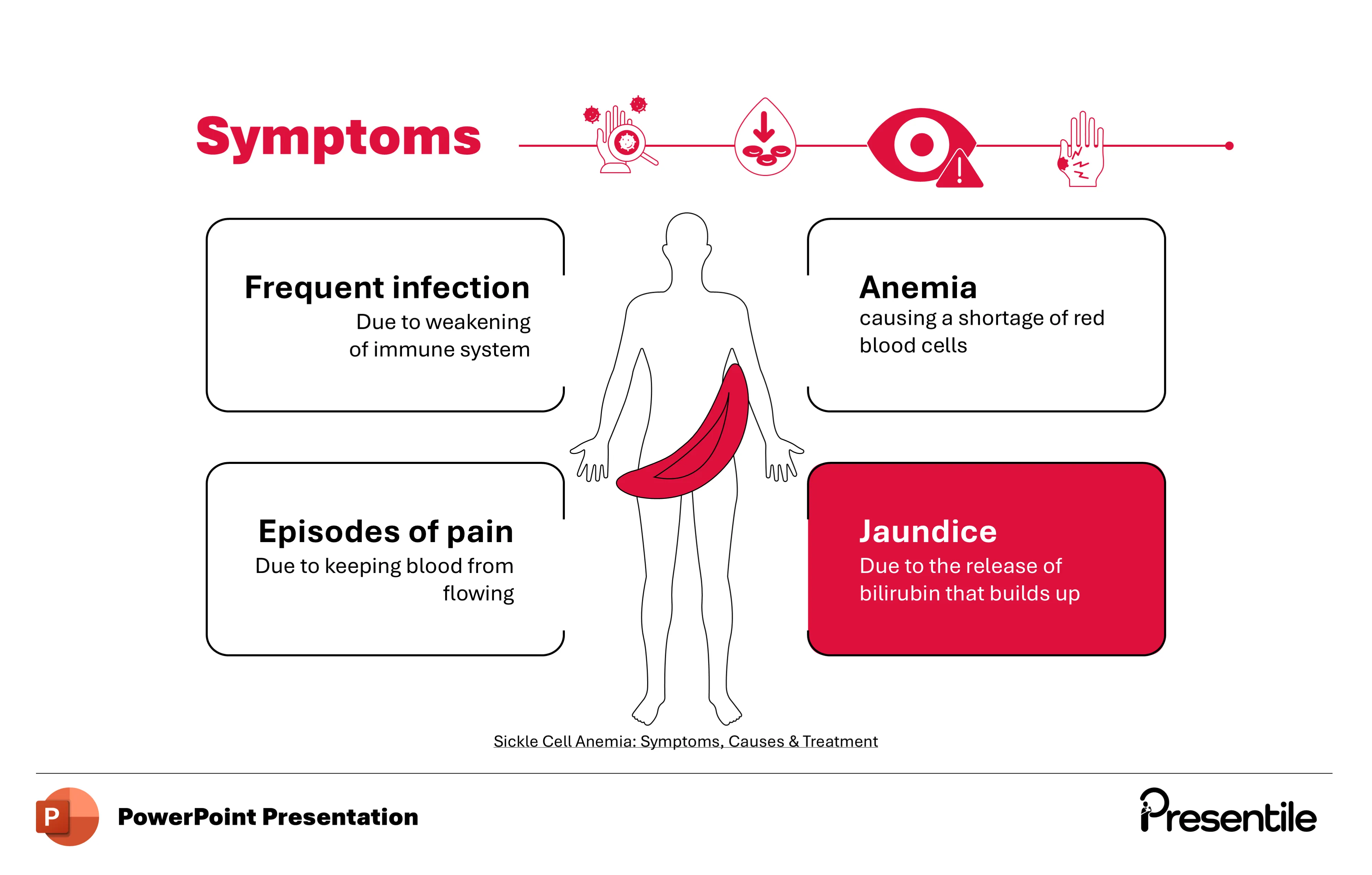
- This slide zeros in on Jaundice as a key symptom of Sickle Cell Anemia.
- The slide uses a bold, red highlight on the Jaundice symptom box to draw attention to it.
- The text explains that Jaundice is caused by the release of bilirubin that builds up in the body.
- This reinforces the interconnectedness of symptoms and pathology, showing that the rapid breakdown of red blood cells (leading to anemia) also releases bilirubin, which in turn causes the yellowing of the skin and eyes characteristic of jaundice.
Slide 10: The Mechanism of Jaundice in Sickle Cell Anemia
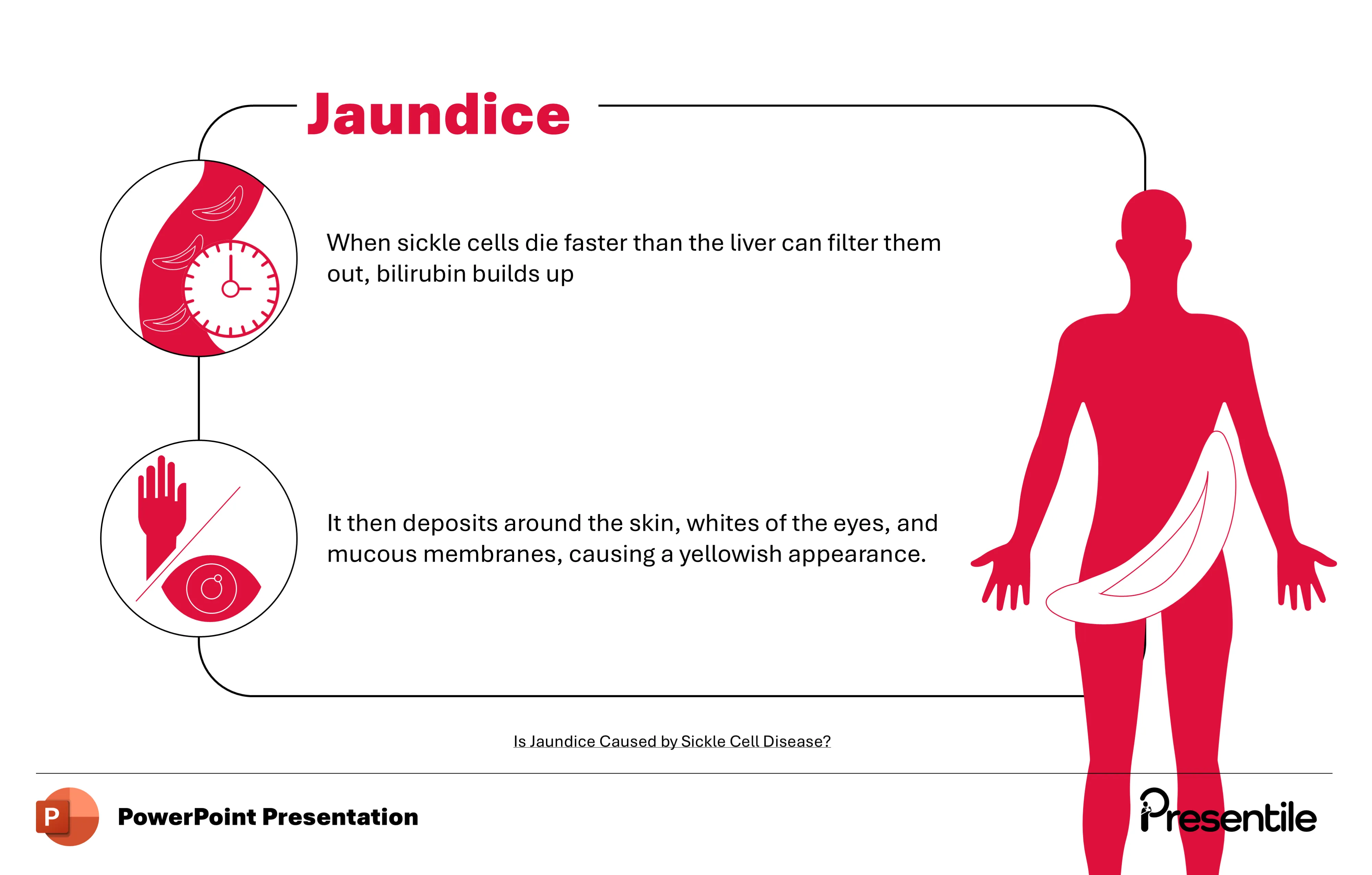
- This slide provides a clear, two-step breakdown of how jaundice develops.
- The first point explains the root cause: when sickle cells die at a rapid rate, the liver is unable to filter out the resulting bilirubin fast enough, leading to its buildup.
- The second point illustrates the consequence of this buildup, showing how bilirubin then deposits in the skin and whites of the eyes, causing the characteristic yellowish appearance.
- This slide effectively connects the cellular pathology to the visible clinical symptom, making a complex process simple to understand.
Slide 11: Highlighting Pain Episodes in Sickle Cell Anemia
- This slide focuses on the symptom of Pain Episodes, a hallmark of Sickle Cell Anemia.
- The design uses a bold, red highlight to draw attention to the "Episodes of pain" symptom box.
- The text concisely explains that the pain is a result of blood being kept from flowing freely.
- This highlights the connection between the physical shape of the sickle cells and the severe pain crises that patients experience, underscoring the most impactful and debilitating symptom of the disease.
Slide 12: The Mechanism of Pain Crises in Sickle Cell Anemia
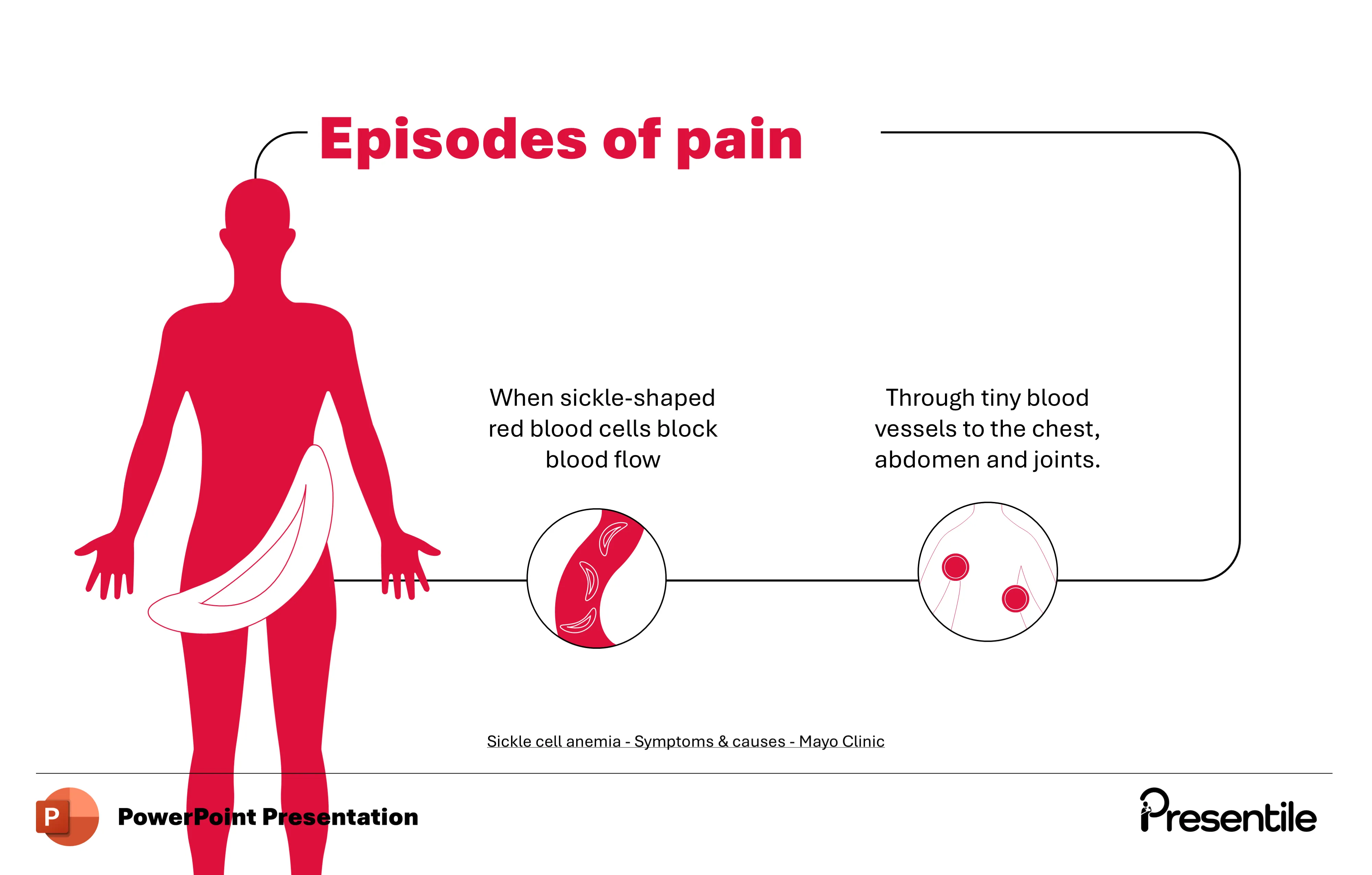
- This slide provides a step-by-step visual explanation of how sickle cells cause pain crises.
- It shows how the abnormal, sickle-shaped red blood cells become lodged in tiny blood vessels, blocking blood flow.
- The slide specifically highlights that this blockage most commonly occurs in the chest, abdomen, and joints, which are the sites of the most intense and frequent pain episodes.
- This clear, anatomical-pathological correlation helps the audience understand the direct cause of the severe pain experienced by patients with the disease.
Slide 13: The Pathophysiology of Sickle Cell Anemia
.webp)
- This slide serves as a powerful introductory slide for the next section on the pathophysiology of Sickle Cell Anemia.
- It uses a bold, clean design with a striking contrast of red and white.
- On the left, a stylized DNA double helix is intertwined with both normal and sickle-shaped red blood cells, visually signaling the genetic cause of the disease.
- The prominent heading "Pathophysiology" clearly prepares the audience for a deep dive into the underlying biological mechanisms. The layout is simple yet impactful, effectively transitioning the presentation to a more detailed, scientific segment.
Slide 14: Comparing Normal vs. Sickle Cells
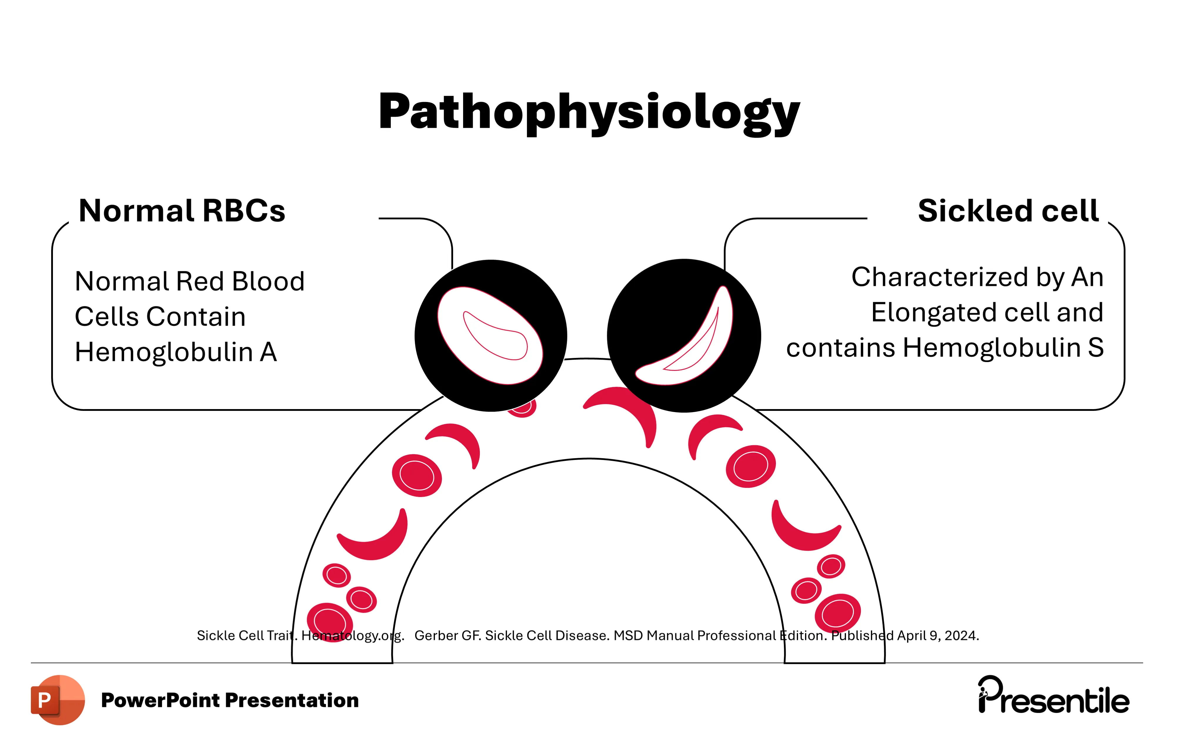
- This slide provides a direct comparison between normal and sickle-shaped red blood cells (RBCs) at the molecular level.
- It clearly labels and contrasts the two types of cells.
- The description for Normal RBCs notes that they contain Hemoglobin A, the standard protein responsible for oxygen transport.
- In contrast, the description for a Sickled cell highlights its elongated shape and states that it contains Hemoglobin S.
- This slide sets the foundation for understanding the genetic and protein-level difference that is the root cause of the disease.
Slide 15: The Genetic Cause of Sickle Cell Anemia
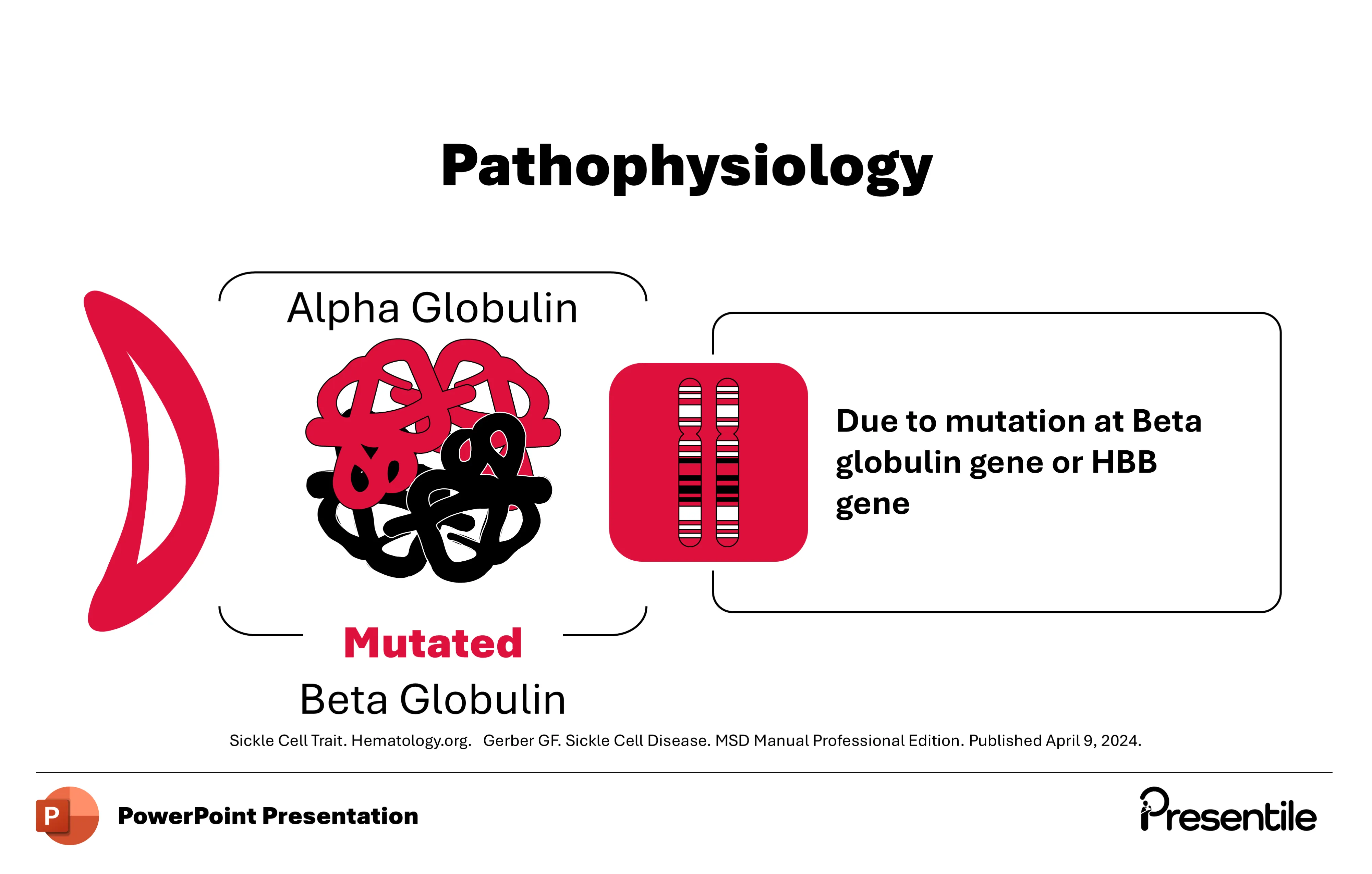
- This slide delves into the specific genetic mutation behind Sickle Cell Anemia.
- It visually links a sickle-shaped cell to the mutated hemoglobin protein.
- The key point is highlighted: the disease is caused by a mutation at the Beta globin gene, also known as the HBB gene.
- This slide effectively simplifies a complex genetic concept, showing how a single gene mutation results in the abnormal beta-globin chain, which in turn leads to the production of sickle hemoglobin and the characteristic cell shape.
- It connects the macroscopic symptom (the sickle cell) to the microscopic genetic cause.
Slide 16: The Polymerization of Hemoglobin S
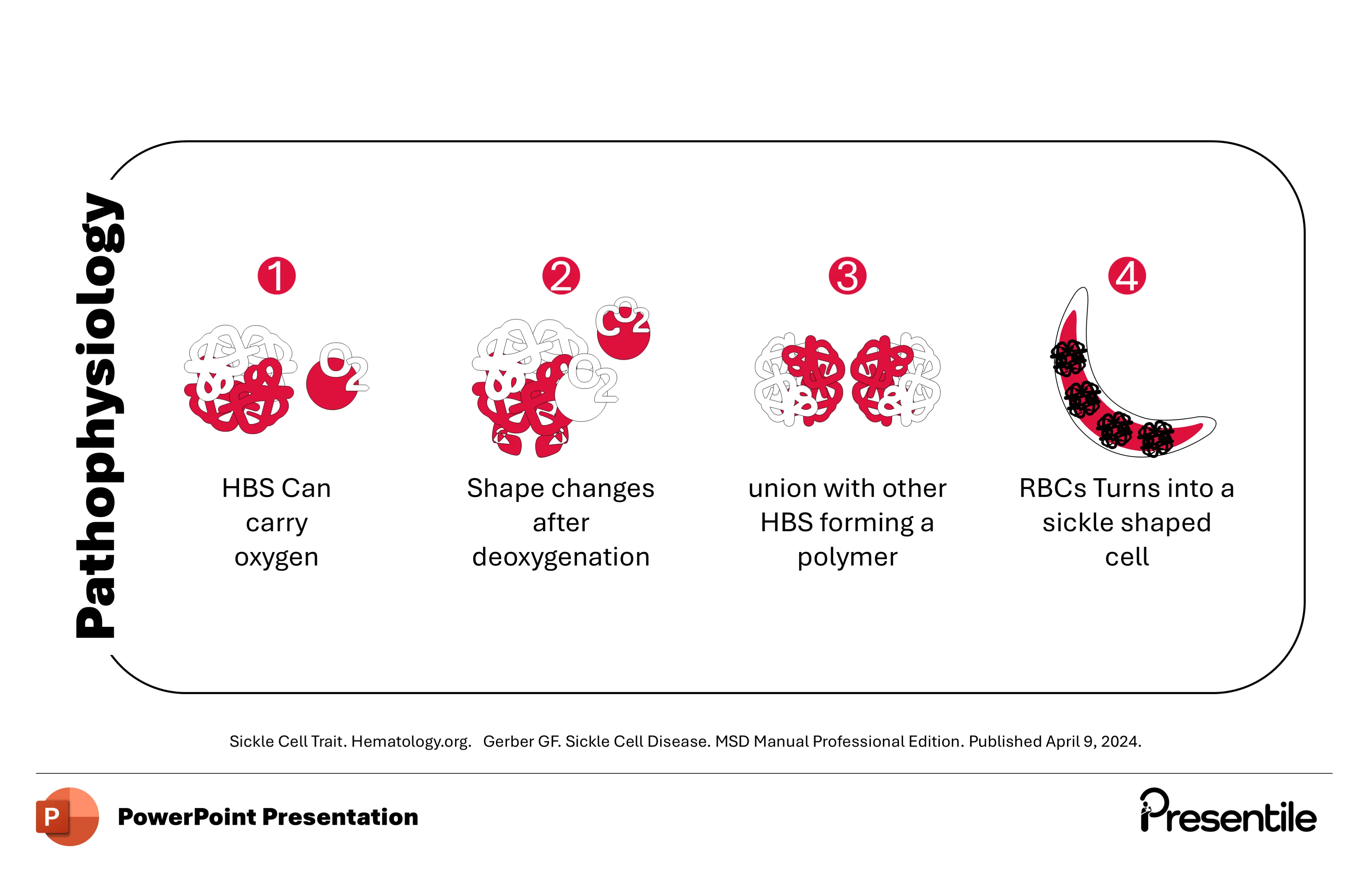
- This slide provides a four-step breakdown of the process that causes red blood cells to sickle.
- It begins with Hemoglobin S (HbS) being able to carry oxygen, just like normal hemoglobin.
- The second step shows that when HbS is deoxygenated, its shape changes.
- The third step illustrates the critical moment: these changed HbS molecules unite to form a long, rigid polymer.
- The final step shows the consequence of this polymerization, as the rigid polymers distort the red blood cell, turning it into the characteristic sickle shape.
- This slide simplifies a complex molecular process, showing a clear cause-and-effect relationship that underpins the entire disease.
Slide 17: The Consequences of Sickling
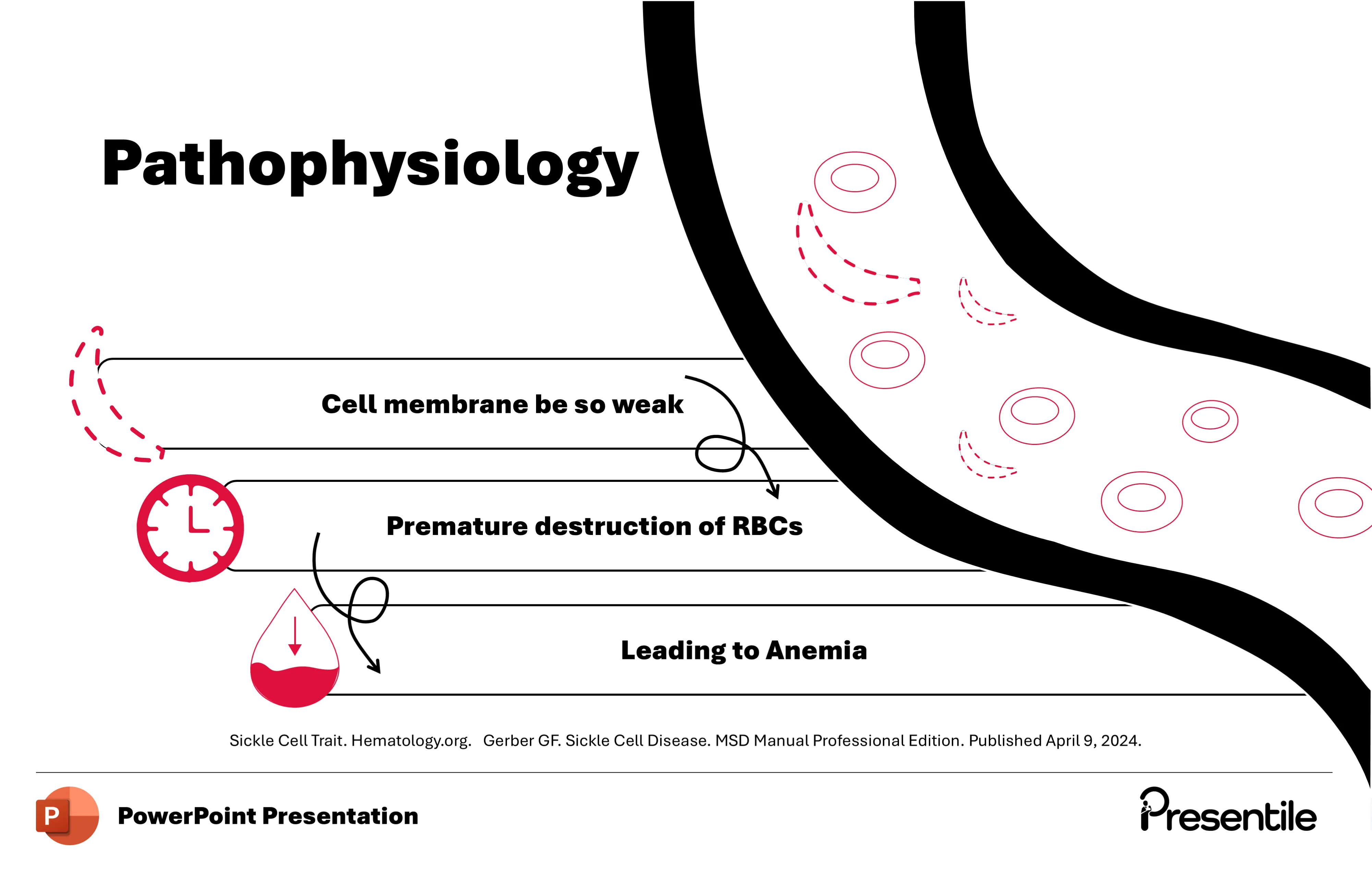
- This slide connects the pathophysiology to the clinical symptoms, showing the domino effect of sickle cell disease.
- It illustrates how the rigid, sickle-shaped red blood cells (RBCs) in the bloodstream have a weakened cell membrane.
- This fragility leads to the premature destruction of the RBCs, a process known as hemolysis, which is much faster than the destruction of normal RBCs.
- The final result of this rapid destruction is a shortage of red blood cells, leading to anemia.
- The visual flow from the sickle cell to its eventual destruction and the resulting anemia provides a clear and concise summary of the disease's core pathological process.
Slide 18: Diagnosis and Genetic Testing
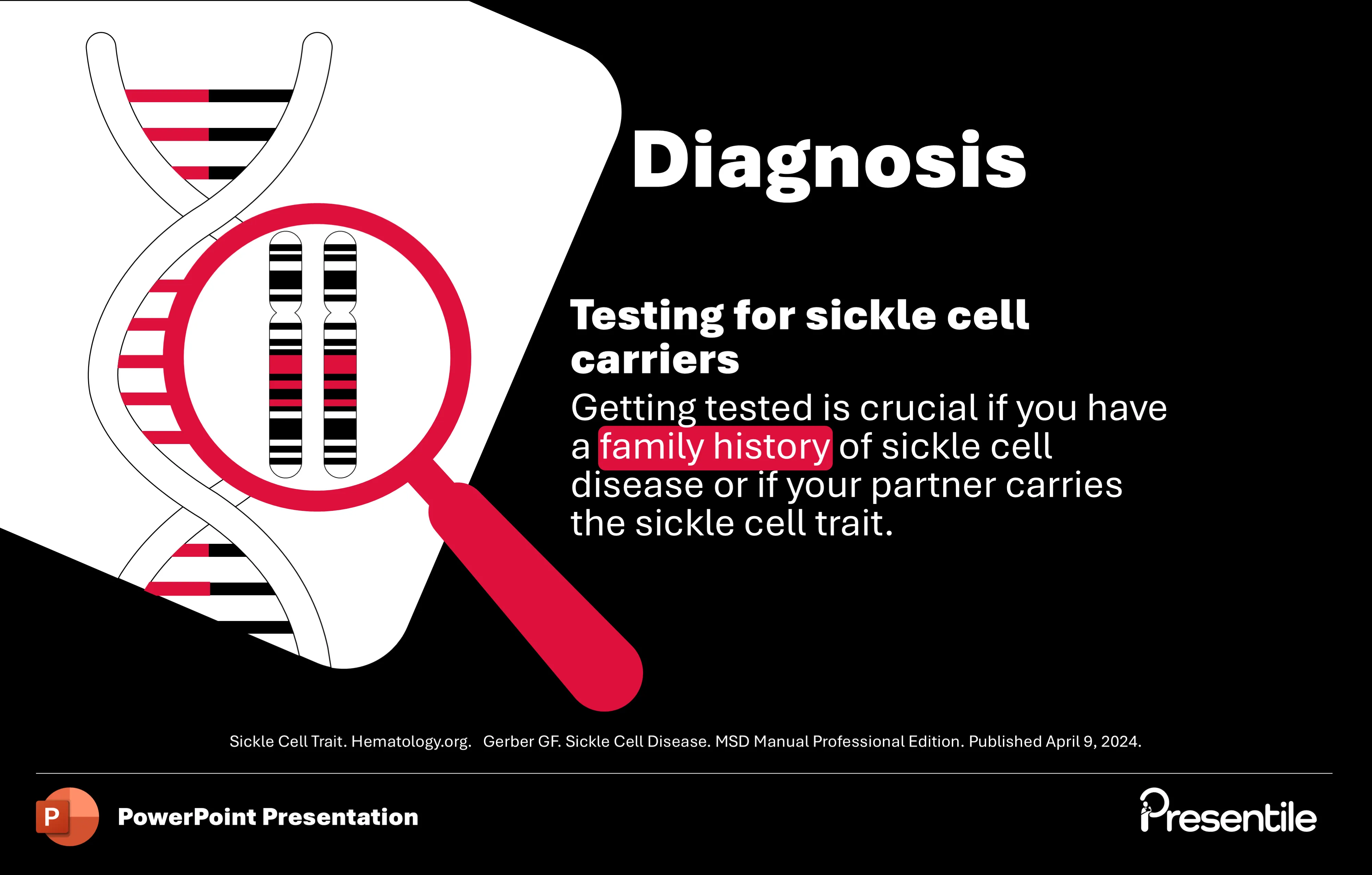
- This slide introduces the diagnostic aspect of Sickle Cell Anemia, with a focus on genetic testing.
- The visual combines a DNA helix and a chromosome under a magnifying glass, emphasizing the genetic nature of the disease.
- The text clearly states that getting tested for sickle cell carriers is crucial, especially for those with a family history or whose partner carries the sickle cell trait.
- This slide highlights the importance of proactive screening and genetic counseling, moving the presentation from a discussion of the disease itself to a practical, preventative consideration for individuals.
Slide 19: Prenatal Screening for Sickle Cell Anemia
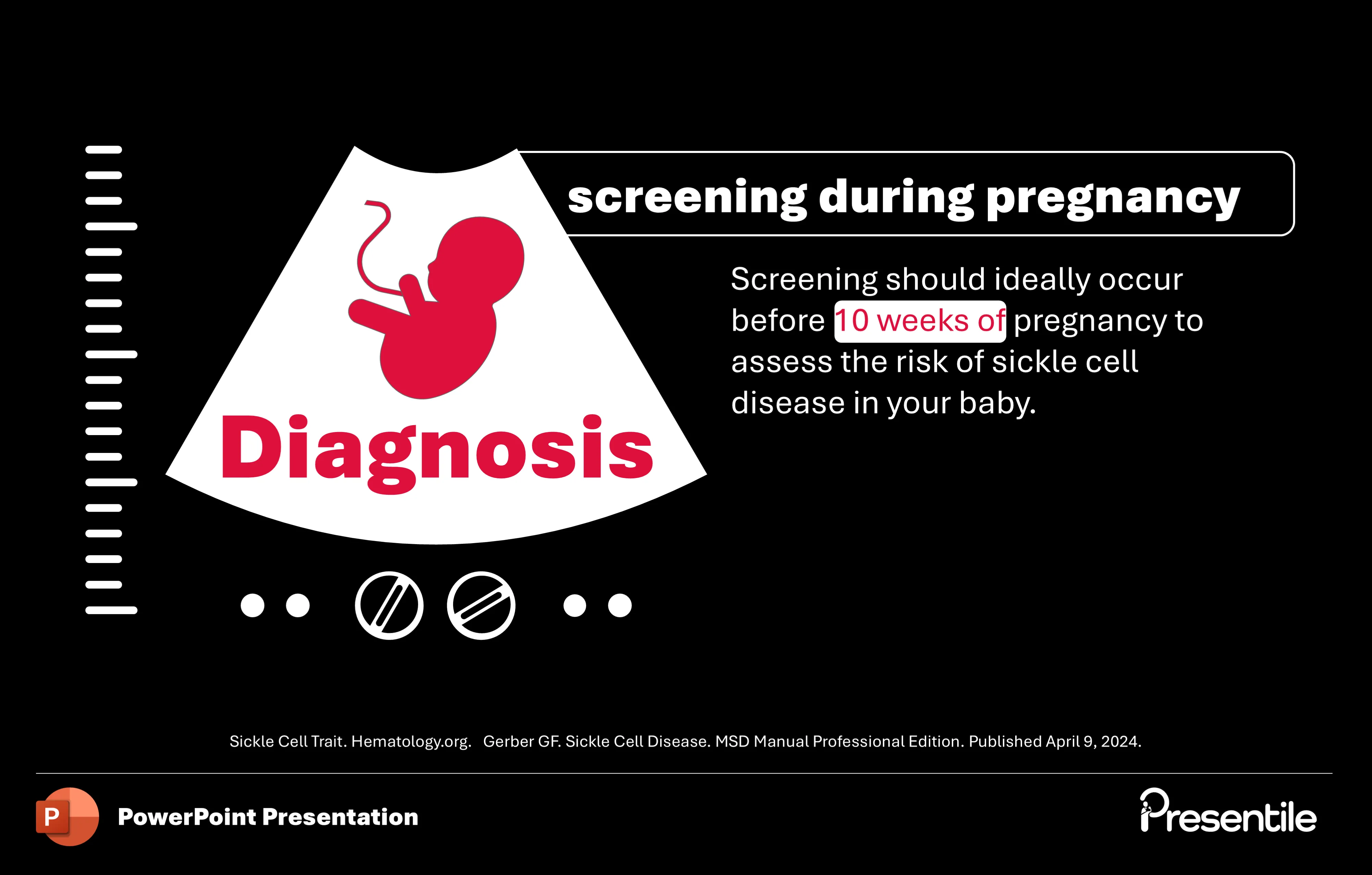
- This slide discusses the importance of screening for Sickle Cell Anemia during pregnancy.
- The visual features an ultrasound graphic with a fetus, immediately setting the context.
- The key message is that screening should ideally occur before 10 weeks of pregnancy to assess the risk of the disease in the baby.
- This slide is crucial for highlighting the preventive and early diagnostic measures available, providing valuable information for expectant parents and healthcare providers on managing genetic risks.
Slide 20: Other Diagnostic Methods
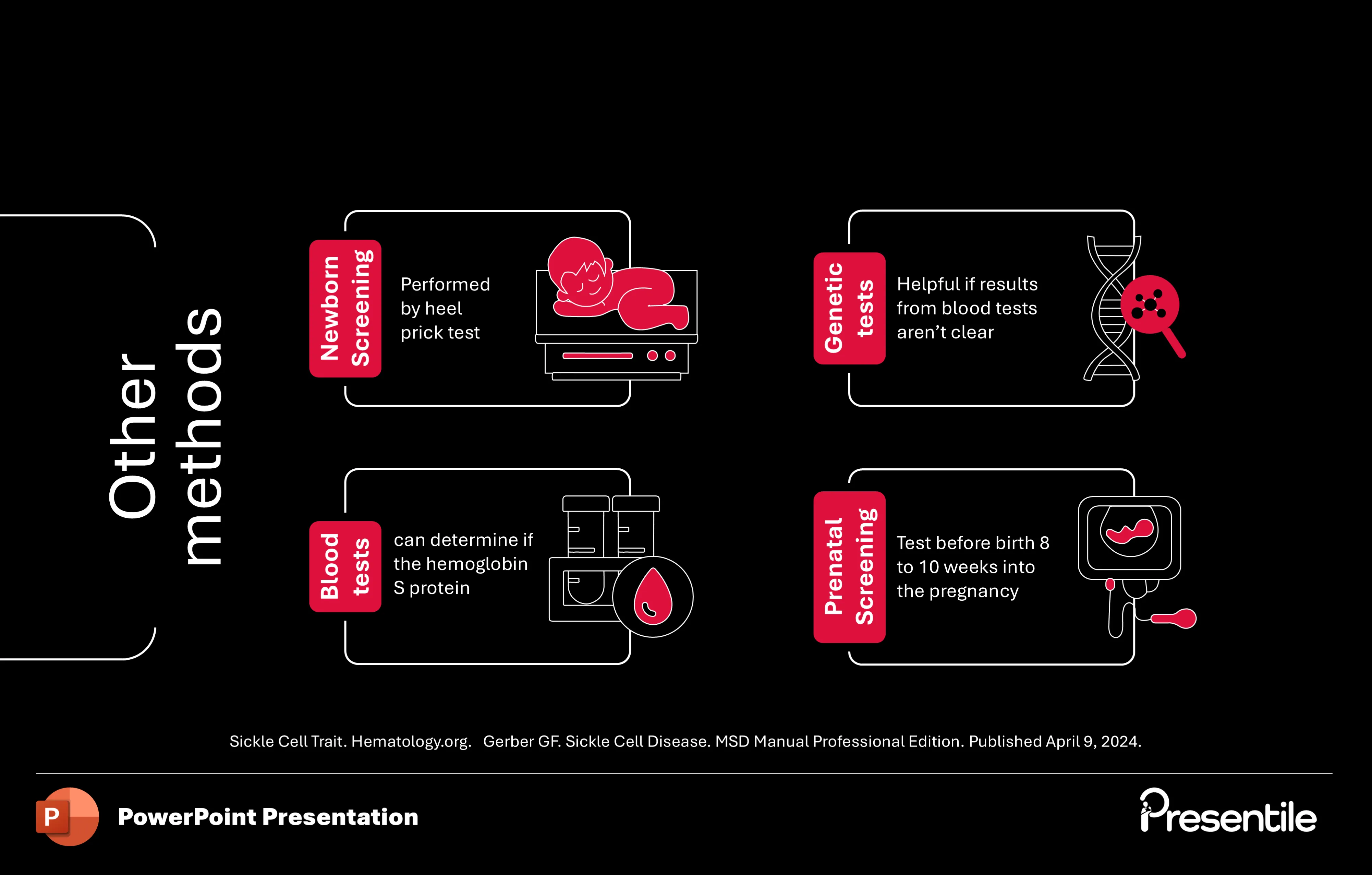
- This slide provides a clear summary of various diagnostic methods for Sickle Cell Anemia.
- It's laid out in a clean, four-quadrant design, with each box dedicated to a different test.
- The methods covered are: Newborn Screening, performed using a heel prick test; Blood tests, which can detect the presence of Hemoglobin S; Genetic tests, used when blood test results are inconclusive; and Prenatal Screening, which is done early in pregnancy.
- This comprehensive overview ensures the audience understands the full range of options available for early detection and diagnosis.
Slide 21: An Overview of Complications in Sickle Cell Anemia
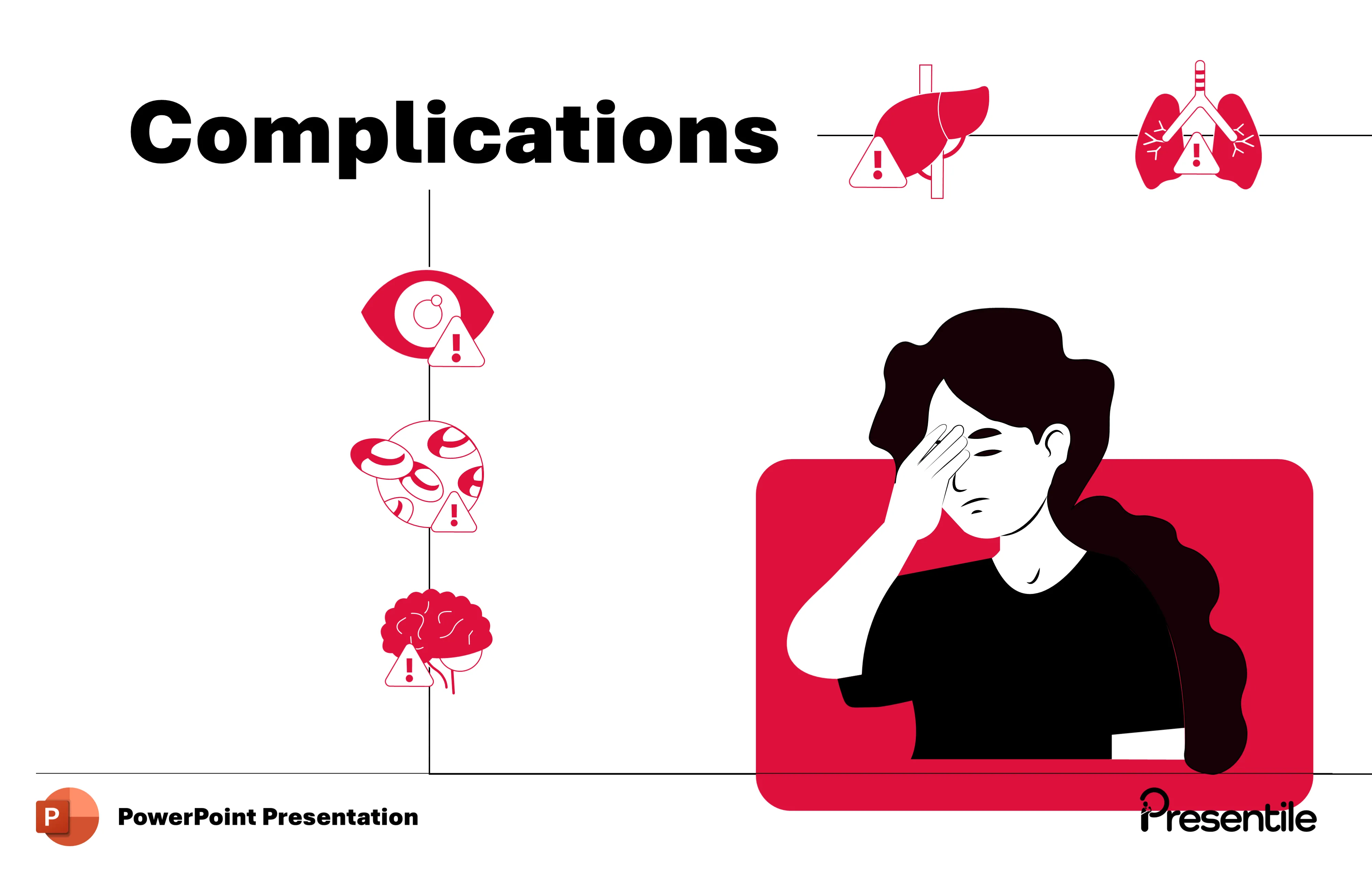
- This slide introduces the critical topic of complications related to Sickle Cell Anemia.
- The design features a prominent title, "Complications," and uses a striking series of icons to visually represent the major systems affected by the disease.
- Icons for the eyes, brain, liver, and lungs are highlighted with warning symbols, immediately drawing attention to the potential risks.
- This slide effectively sets the stage for a detailed discussion of the serious, long-term health issues that can arise from the disease.
Slide 22: Complications: Leg Ulcers, Vision Damage, and Acute Chest Syndrome

- This slide details three significant complications of Sickle Cell Anemia. It uses a clean, three-panel layout with distinct visuals for each.
- The first panel describes Leg Ulcers, explaining that painful open sores can form on the legs.
- The second panel covers Vision Damage, noting that sickle cells can block tiny blood vessels in the eyes.
- The third and final panel highlights Acute Chest Syndrome, a serious condition that involves a lung infection or sickle cells blocking blood vessels in the lungs.
- This slide provides a clear and concise summary of these severe health consequences, reinforcing the gravity of the disease.
Slide 23: Stem Cell Transplantation as a Treatment
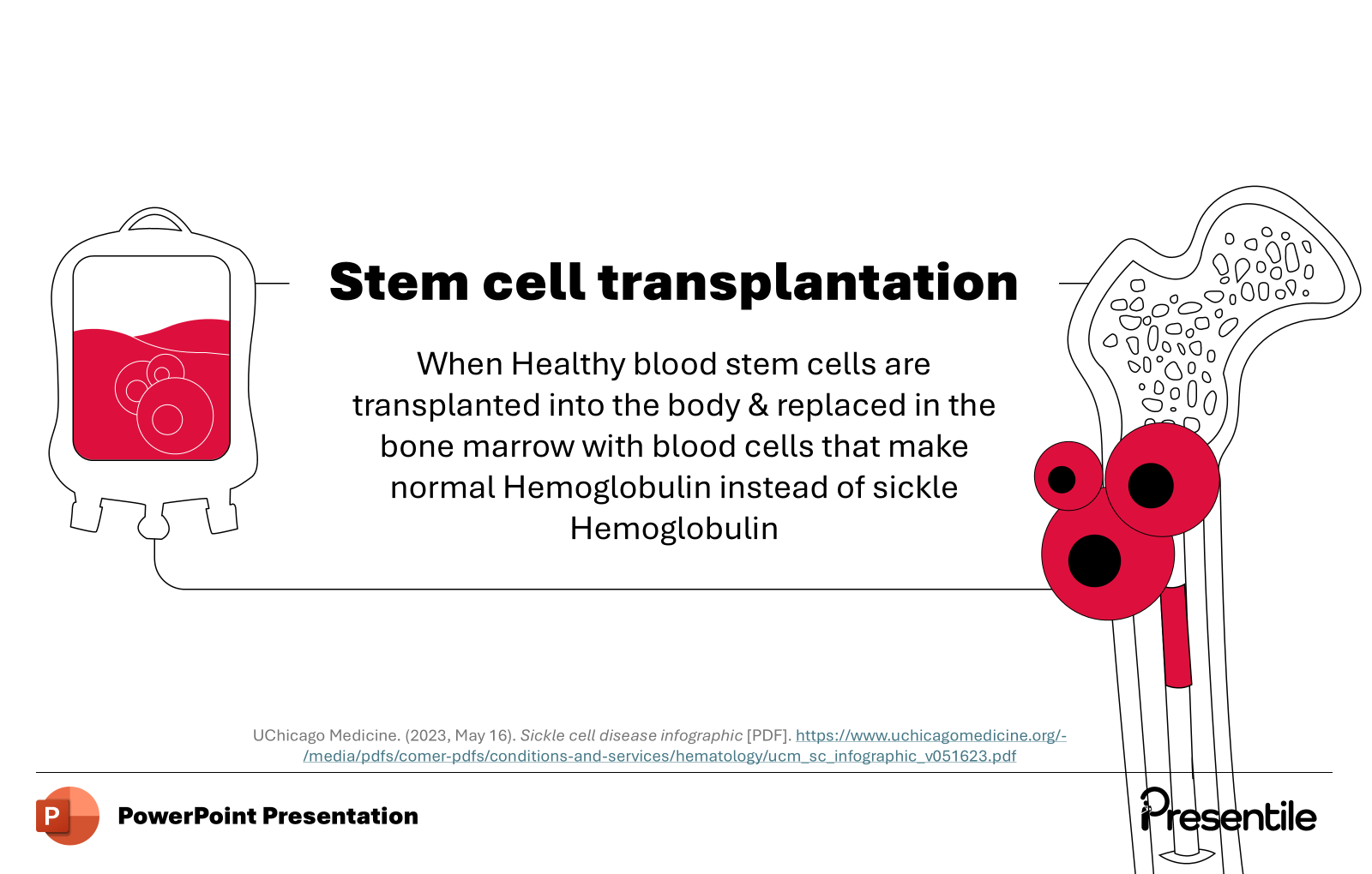
- This slide introduces stem cell transplantation as a major treatment for Sickle Cell Anemia.
- The design features a clear visual of an IV bag connected to a stylized bone, representing the transfer of healthy blood cells into the bone marrow.
- The description explains the core principle of the treatment: healthy blood stem cells are transplanted into the body to replace the bone marrow cells that produce sickle hemoglobin.
- This provides a clear, high-level understanding of this potentially curative but complex therapeutic option.
Slide 24: The Process of Stem Cell Transplantation
- This slide provides a clear, step-by-step breakdown of the stem cell transplantation process.
- It begins by showing how blood is collected from a donor. The next step illustrates the crucial process of separating the stem cells from the donor's blood.
- The final step shows how these donor stem cells are infused into the patient, typically via a catheter.
- This simple, linear flow diagram makes a complex medical procedure easy to understand, highlighting the key stages from donation to infusion.
Slide 25: Treatment Options for Sickle Cell Anemia
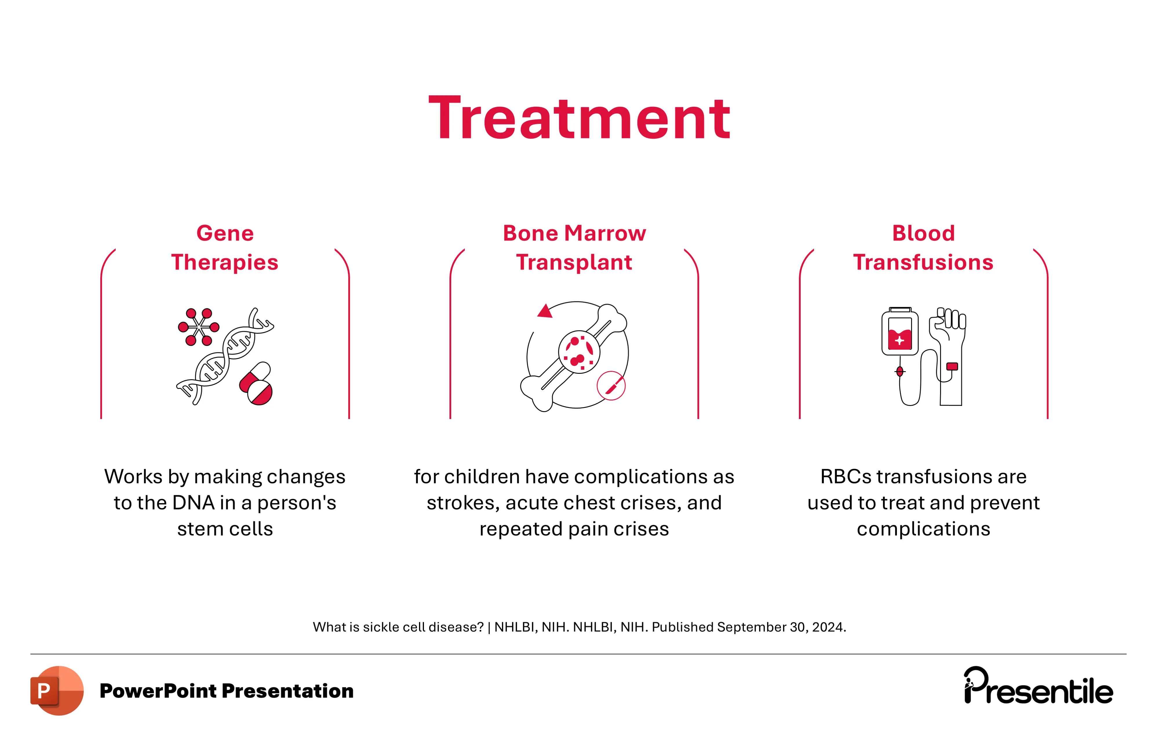
- This slide provides a comprehensive overview of the three primary treatment options for Sickle Cell Anemia.
- It uses a clean, three-column layout, with each column dedicated to a different therapy.
- The first column describes Gene Therapies, explaining that they work by making changes to the DNA in a patient's stem cells.
- The second column focuses on Bone Marrow Transplants, noting their use in children with severe complications like strokes or acute chest crises.
- The final column covers Blood Transfusions, a common treatment used to manage and prevent complications.
- This slide serves as an excellent summary of the available therapeutic strategies, from the most advanced to the most common.
Slide 26: Thank You and Conclusion
- This slide serves as a professional and clean conclusion to the presentation.
- The "Thank you" is displayed in large, bold text, providing a clear final message to the audience.
- The stylized sickle cell graphic on the left ties the final slide back to the presentation's core topic, maintaining a cohesive design.
- This slide is a simple yet effective way to end the presentation and can be used to open the floor for questions or a final summary.
Features of
Sickle Cell Anemia Presentation Monochrome
- Fully editable in PowerPoint
- All graphics are in vector format
- Medically Referenced information and data
Specifications
 Slides count:
 Compatible with:Microsoft PowerPoint
 File type:PPTX
 Dimensions:16:9
Files Included
- Non-animated PowerPoint
- Animated PowerPoint File
- Animated PowerPoint with Voice Over
- PDF Documents with presentation script
Elevate Your Work with Our Innovative Slides
Thank you! Your submission has been received!
Oops! Something went wrong while submitting the form.
No items found.