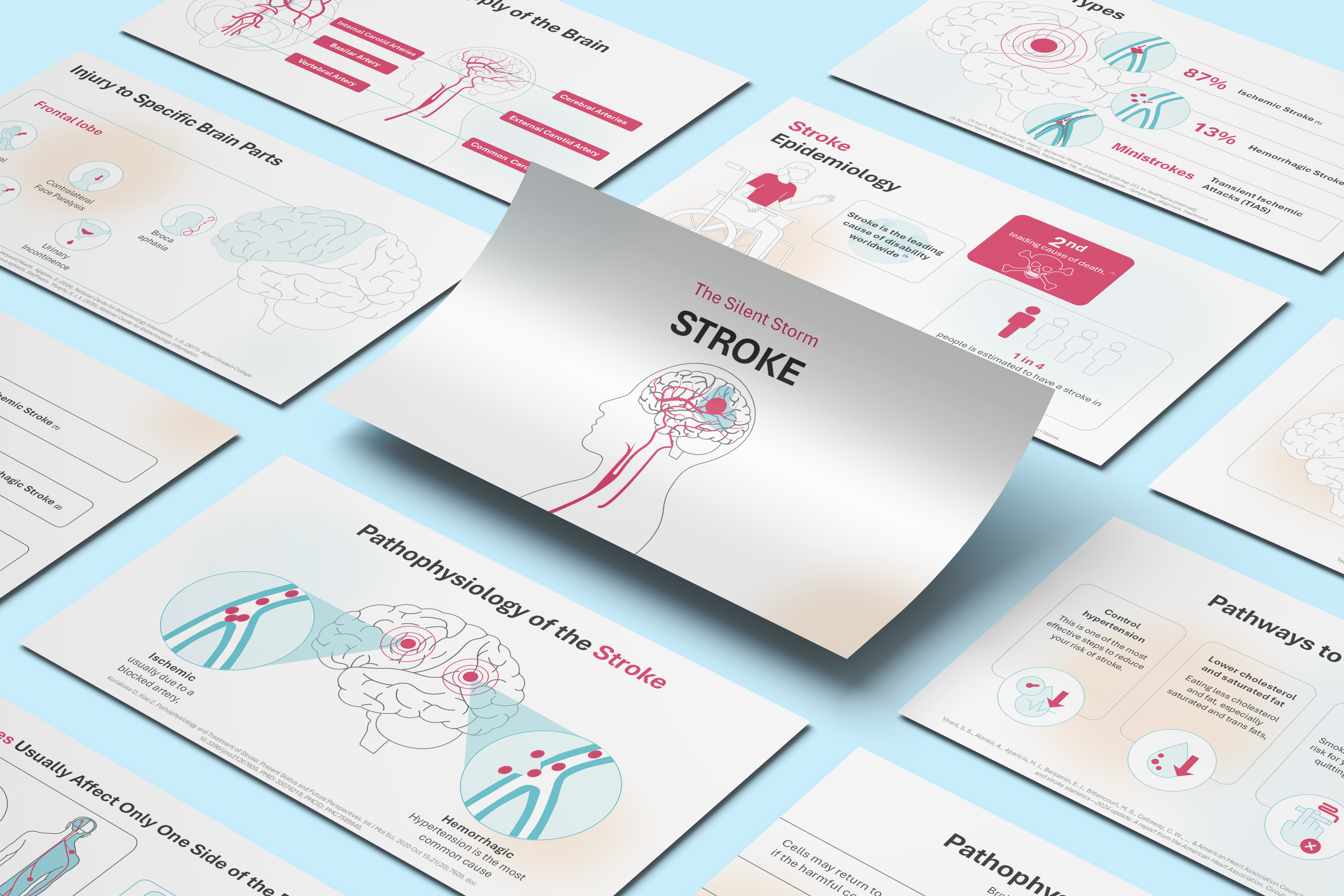
This powerful title slide introduces a presentation focused on the serious cerebrovascular event: Stroke.
The design is impactful, using the phrase "The Silent Storm" as a dramatic subtitle.
Main Title: The word STROKE is prominently displayed in bold red and black text.
Visual: The central graphic is a simplified side-view of a human head and brain, showing the carotid artery and cerebral blood vessels. A red area and blue shading clearly illustrate an event—likely a hemorrhagic or ischemic block—in the brain's vascular structure, visually conveying the critical nature of the topic.
Slide 2: Brain Anatomy
Brain Anatomy
This slide provides a clear, labeled illustration of the major regions of the human brain, which is essential context for understanding how different stroke locations affect function.
The graphic shows a lateral view of the brain with the following lobes/regions clearly labeled in red banners:
Frontal lobe
Parietal lobe
Temporal lobe
Occipital lobe
Cerebellum
Brain stem
This slide prepares the audience by establishing the relationship between brain anatomy and the subsequent discussion of stroke location and corresponding symptoms (localization of function).
Slide 3: Blood Supply of the Brain (Cerebral Vasculature)
Blood Supply of the Brain (Cerebral Vasculature)
This slide provides a detailed look at the major arterial structures that supply blood to the brain, which is the direct focus of stroke pathophysiology. The visual presents two views: a frontal view of the entire brain vasculature and a lateral view showing the origin of the vessels.
Key structures labeled are:
Common Carotid Artery
External Carotid Artery (Supplying the face/neck, less relevant to cerebral stroke)
Internal Carotid Arteries (Major supply to the brain)
Vertebral Artery (Major supply to the posterior circulation)
Basilar Artery (Formed by the fusion of the vertebral arteries)
Cerebral Arteries (The main branches within the brain)
Circle of Willis (The crucial anastomotic ring at the base of the brain)
Slide 4: Definition of Stroke
Definition of Stroke
This slide provides a clear, concise definition of a stroke, linking it directly back to the visual established on the title slide.
Question: "What is Stroke?" is asked in a prominent red banner, signaling the definition.
Definition: The stroke is defined as being "Caused by blocked blood flow to the brain or sudden bleeding in the brain." This encompasses the two main types of stroke: ischemic (blocked flow) and hemorrhagic (bleeding).
The same powerful graphic of the brain's blood supply with the localized event is used, reinforcing the concept of a disruption to cerebral circulation.
Slide 5: Stroke Epidemiology
Stroke Epidemiology
This slide transitions the focus to the global burden and statistical impact of stroke. The title is clear, and the slide uses three powerful statistics to convey the seriousness of the disease.
Key Statistic 1 (Disability): "Stroke is the leading cause of disability worldwide." This highlights the profound long-term impact on quality of life, reinforced by the visual of a person in a wheelchair.
Key Statistic 2 (Mortality): "2nd leading cause of death." This emphasizes the high fatality rate associated with stroke, symbolized by the skull and crossbones icon.
Key Statistic 3 (Lifetime Risk): "1 in 4 people is estimated to have a stroke in their lifetime." This conveys the high prevalence and broad risk across the general population.
Slide 6: Stroke Types
Stroke Types
This crucial slide classifies the three main types of cerebrovascular events, detailing the prevalence of each. The central visual shows a brain with a warning signal, and three magnified artery diagrams clearly illustrate the pathology of each type.
Ischemic Stroke:
Prevalence: 87% of all strokes.
Pathology: Caused by a blockage (a clot or embolus) in a cerebral artery, cutting off blood supply.
Hemorrhagic Stroke:
Prevalence: 13% of all strokes.
Pathology: Caused by a ruptured blood vessel that bleeds into the brain tissue or surrounding spaces.
Transient Ischemic Attacks (TIAs) / Ministroke:
Pathology: A temporary blockage with symptoms lasting less than 24 hours (usually minutes), often serving as a warning sign for a full stroke.
Slide 7: Pathophysiology of the Stroke
Pathophysiology of the Stroke
This slide serves as the transition into the detailed mechanisms of stroke (pathophysiology), linking the two main types of stroke to their root causes. The central image of the brain with concentric red circles reinforces the area of injury.
Ischemic Stroke:
Cause: "Ischemic usually due to a blocked artery." This re-emphasizes that lack of blood flow due to an occlusion is the primary mechanism.
Visual: A magnified view of an artery with a blockage (likely a thrombus or embolus) cutting off flow.
Hemorrhagic Stroke:
Cause: "Hypertension is the most common cause." This identifies the leading risk factor and mechanism (high pressure leading to rupture) for bleeding strokes.
Visual: A magnified view of an artery showing leakage or rupture, leading to extravasated blood cells.
Slide 8: Pathophysiology of the Stroke – Duration and Damage
Pathophysiology of the Stroke – Duration and Damage
This slide provides critical information about the time-dependent nature of stroke damage, emphasizing the urgency of treatment. The central image of the affected brain and the stopwatch visually reinforce the message.
Key Concept: "Brain damage depends on blood loss duration." This is the core message of "time is brain."
Short Duration: "Cells may return to normal function if the harmful condition is brief." This refers to the penumbra, the salvageable brain tissue surrounding the core infarct.
Prolonged Duration: "But die if prolonged. Some die in minutes, others after 30+ minutes." This highlights the rapid, permanent destruction of tissue in the ischemic core.
Slide 9: Risk Factors for Stroke
Risk Factors for Stroke
This slide details five major modifiable and non-modifiable risk factors for stroke. It maintains the visual theme, featuring the brain with the area of injury on the left, and lists the risk factors clearly on the right.
High blood pressure: A critical and controllable factor in stroke (especially hemorrhagic).
Diabetes: Increases the risk of atherosclerosis, leading to ischemic stroke.
Unhealthy diet: Contributes to conditions like high cholesterol and hypertension.
Smoking and alcohol: Major lifestyle choices that significantly increase stroke risk.
Obstructive sleep apnea: A sleep disorder increasingly recognized as an independent stroke risk factor.
Slide 10: Risk Factors for Stroke (Continued)
Risk Factors for Stroke (Continued)
This slide continues the discussion on risk factors, focusing on vascular, cardiac, and lifestyle risks not covered on the previous slide. It maintains the clear structure with the brain visual on the left.
High cholesterol levels: Contributes to atherosclerosis, a major precursor to ischemic stroke.
Heart disorders: Conditions like Atrial Fibrillation (AFib) are a major source of emboli that can cause ischemic stroke.
Obesity: A systemic condition that contributes to hypertension, diabetes, and heart disease—all major stroke risk factors.
Lack of physical activity: A major modifiable lifestyle factor that increases the risk of cardiovascular disease.
Use of cocaine/amphetamines: These substances can cause sudden, severe hypertension and arterial spasm, leading to hemorrhagic or ischemic stroke.
Slide 11: Symptoms of Stroke – Hemiparesis and Sensory Loss
Symptoms of Stroke – Hemiparesis and Sensory Loss
This crucial slide introduces the key signs and symptoms of a stroke, emphasizing the characteristic lateralization of effects. The central graphic is highly informative, showing the pathway from brain lesion to physical manifestation.
Key Concept: "Strokes Usually Affect Only One Side of the Body." The visual shows a lesion in one hemisphere of the brain, leading to deficits on the opposite side of the body.
Core Symptoms: The slide lists six common acute symptoms:
Sudden weakness or paralysis (Hemiparesis/Hemiplegia)
Sudden loss of sensation
Sudden difficulty speaking (Dysarthria/Aphasia)
Sudden confusion
Sudden dimness (Vision loss)
Sudden dizziness (Vertigo/Loss of balance)
Slide 12: Injury to Specific Brain Parts (Frontal Lobe)
Injury to Specific Brain Parts (Frontal Lobe)
This slide connects the anatomical concepts from earlier slides to the functional deficits observed in a stroke, specifically focusing on the Frontal Lobe. The visual highlights the frontal lobe area on the brain graphic.
The slide details five common deficits resulting from a frontal lobe stroke:
Contralateral Arm Paralysis: Weakness or inability to move the arm on the opposite side of the body.
Contralateral Face Paralysis: Weakness or drooping of the face on the opposite side.
Broca aphasia: Difficulty in producing speech (expressive aphasia).
Behavior Alteration: Changes in personality, judgment, and emotional control.
Urinary Incontinence: Loss of bladder control, often due to damage to executive control centers.
Slide 13: Injury to Specific Brain Parts (Parietal, Temporal, and Occipital Lobes)
Injury to Specific Brain Parts (Parietal, Temporal, and Occipital Lobes)
This slide continues the discussion on neurological localization, detailing the functional deficits caused by stroke in the Parietal, Temporal, and Occipital lobes. The visual highlights these regions on the brain graphic.
The slide details three key functional deficits:
Parietal Lobe:
Spatial Neglect: Inability to pay attention to one side of space (often the side opposite the lesion).
Wernicke Aphasia: Difficulty understanding language (receptive aphasia).
Temporal Lobe:
Auditory Agnosia: Inability to recognize or distinguish sounds.
Occipital Lobe:
Visual Agnosia: Inability to recognize or interpret familiar objects through sight.
Slide 14: Stroke Warning Signs - The B.E. F.A.S.T. Acronym
Stroke Warning Signs - The B.E. F.A.S.T. Acronym
This critical slide introduces the well-known acronym used by emergency services to quickly identify and respond to stroke victims. The visual shows the acronym components arranged in circles with the affected brain above.
Slide 15: B.E. F.A.S.T. Acronym Defined
B.E. F.A.S.T. Acronym Defined
This slide provides the full definition of the B.E. F.A.S.T. acronym, acting as the critical tool for recognizing and responding to a stroke. The acronym letters are arranged around the affected brain visual, directly connecting the warning signs to the organ in distress.
The acronym breaks down the key symptoms and the necessary response:
B - Balance: Loss of balance or coordination.
E - Eyes: Changes in vision.
F - Face: Drooping features on one side of the face.
A - Arms: Weakness in a limb.
S - Speech: Difficulty speaking or understanding others.
T - Time: Call 911 or emergency services right away.
Slide 16: Stroke Complications
Stroke Complications
This slide transitions the focus from acute stroke recognition to the potential long-term and immediate adverse outcomes (complications) following a stroke. It emphasizes that complications depend on the duration of blood loss and the brain region affected.
Core Message: A stroke can sometimes cause temporary or permanent disabilities. Complications depend on how long the brain lacks blood flow and which part is affected.
Specific Complications: The slide lists four key complications:
Raised pressure leads to brain herniation: This is a severe and often fatal complication of swelling (edema) after a stroke.
Pressure Sores: A complication often related to immobility following stroke.
Aspiration Pneumonia: A common complication related to difficulty swallowing (dysphagia).
Muscle Loss: Also known as atrophy, related to lack of use and immobility.
Slide 17: Stroke Complications (Continued)
Stroke Complications (Continued)
This slide continues the list of potential adverse outcomes following a stroke, focusing on cardiovascular and infectious complications. It maintains the visual theme of categorized complications.
Pulmonary Embolism (PE): A serious complication where a blood clot (often from Deep Vein Thrombosis) travels to the lungs.
Deep Vein Thrombosis (DVT): Blood clot formation, usually in the leg, due to immobility following a stroke.
Urinary Tract Infections (UTI): Common complications related to catheter use or bladder dysfunction after a stroke.
Permanent Shortening of Muscles: This refers to contractures, a severe form of muscle stiffness and shortening caused by prolonged lack of use and abnormal muscle tone.
Slide 18: Unmasking Stroke: The Diagnostic Path
Unmasking Stroke: The Diagnostic Path
This slide outlines the three key steps in diagnosing a stroke, emphasizing the tools used to distinguish between ischemic and hemorrhagic types. The visual shows a patient undergoing a diagnostic scan.
Medical History and Examination: Doctors assess strength, reflexes, speech, vision, balance, and medical history. This initial step utilizes the B.E. F.A.S.T. signs (from Slide 15) and other neurological checks.
CT Scan (Computed Tomography): This is the fastest tool to detect bleeding and rule out hemorrhagic stroke. Rapid CT is essential because treatment differs drastically based on the stroke type.
MRI (Magnetic Resonance Imaging): Provides more detailed images, useful for identifying smaller or earlier strokes. This is often used to confirm the diagnosis and determine the extent of damage.
Slide 19: Treatment of Stroke
Treatment of Stroke
This slide provides a high-level overview of the five categories of intervention used in the acute and chronic management of stroke. The visual uses five icons in a circuit pattern, suggesting a multi-faceted and continuous approach to care.
Vital Support: Focuses on stabilizing the patient's immediate condition, likely including airway, breathing, and circulation management.
Clot Treatment: Refers to thrombolytic drugs (like tPA) used to dissolve clots in ischemic stroke.
Endovascular Therapy: Refers to mechanical thrombectomy, a procedure to physically remove a large clot from a blocked artery.
Rehabilitation Therapy: Involves physical, occupational, and speech therapy to regain function after brain damage.
Post-Stroke Treatment: Encompasses long-term management of risk factors (like blood pressure) and prevention of recurrence.
Slide 20: Pathways to Prevent Stroke
Pathways to Prevent Stroke
This slide transitions the focus to the long-term, preventative strategies essential for reducing the risk of a stroke. This information serves as a powerful call to action based on the risk factors discussed earlier.
The slide details four core preventative measures:
Control hypertension: This is one of the most effective steps to reduce your risk of stroke.
Lower cholesterol and saturated fat: Eating less cholesterol and fat, especially saturated and trans fats, helps to lower risk.
Quit tobacco: Smoking raises the risk for you and others; quitting lowers it.
Maintain a healthy weight: Overweight raises risks like hypertension, heart disease, and diabetes.
Slide 21: Preventive Medicines
Preventive Medicines
This slide details the two main classes of medications used to prevent strokes, particularly focusing on preventing ischemic stroke recurrence. The slide is divided into two clear sections:
Antiplatelet Medications:
Function: Anti-platelet medicines make blood cells less sticky and less likely to clot.
Example: The most commonly used anti-platelet medicine is aspirin.
Anticoagulants:
Function: These medicines reduce blood clotting.
Example: Heparin is a fast-acting anticoagulant that may be used short-term in the hospital.
Slide 22: Thank You (Conclusion)
Thank You (Conclusion)
This slide serves as the concise and direct conclusion to your comprehensive 22-slide presentation on Stroke.
The large, bold text "Thank You" formally ends the content portion of the presentation, signaling the audience for a question and answer period. The minimalist design focuses attention on the final message.
Lorem ipsum dolor sit amet, consectetur adipiscing elit. Suspendisse varius enim in eros elementum tristique. Duis cursus, mi quis viverra ornare, eros dolor interdum nulla, ut commodo diam libero vitae erat. Aenean faucibus nibh et justo cursus id rutrum lorem imperdiet. Nunc ut sem vitae risus tristique posuere.
Features
This is some text inside of a div block.
Elevate Your Work with Our Innovative Slides
Thank you! Your submission has been received!
Oops! Something went wrong while submitting the form.