Medical Presentations
Respiratory
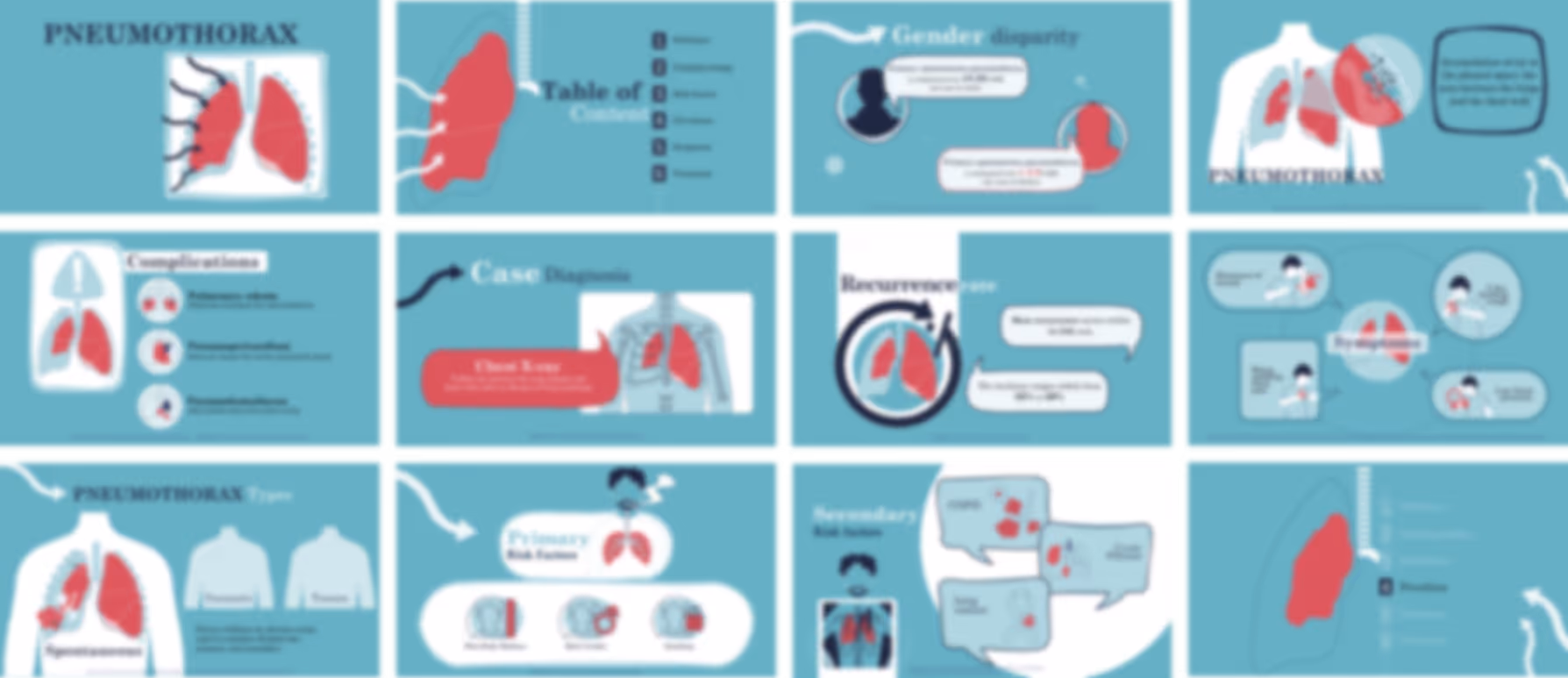
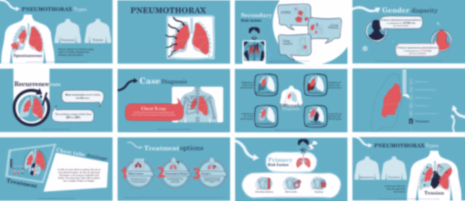
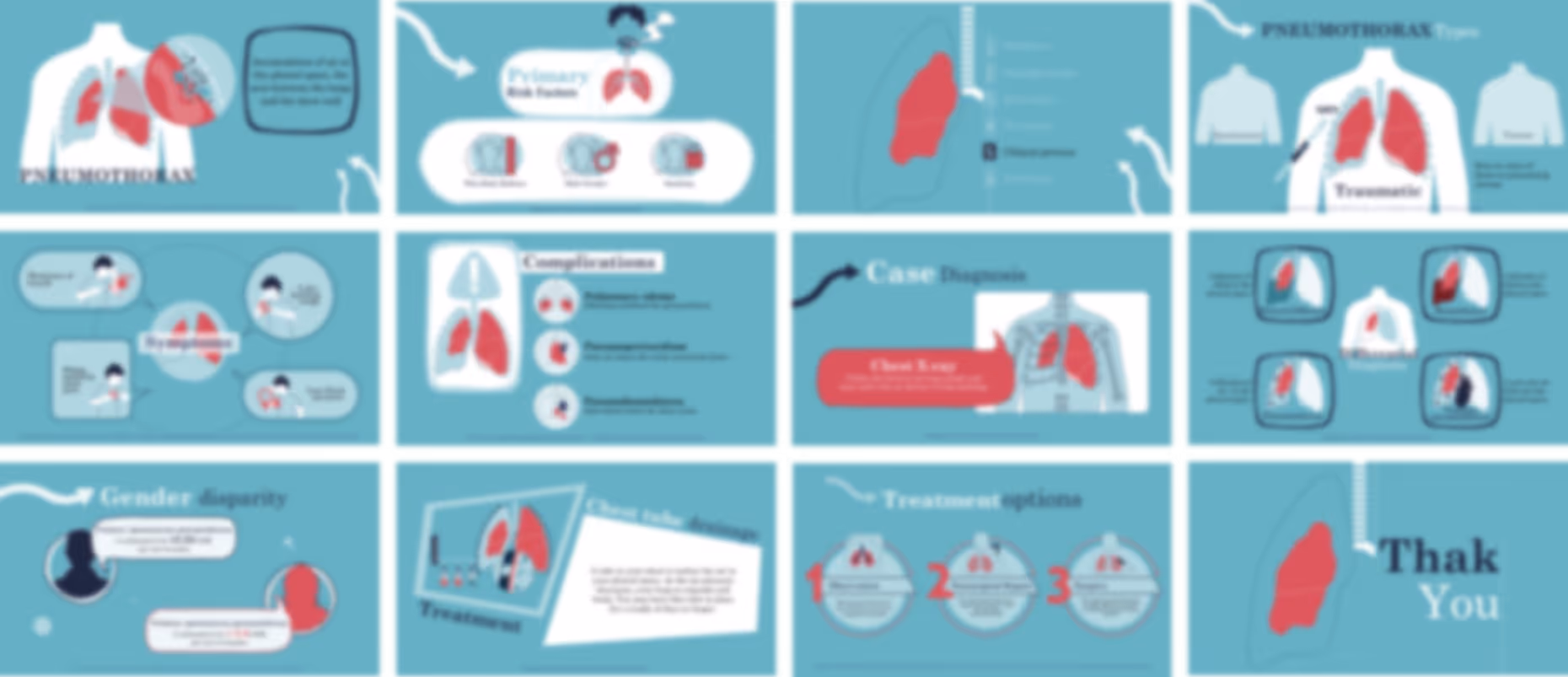
Pneumothorax Overview Presentation

No items found.
Trusted By Life Scinece Teams:


Fully editable in PowerPoint with customizable colors, shapes, and layouts
For easy and friendly drag and drop
Fully animated slides
To help simplify and communicate complex medical concepts
All graphics in vector format
For crisp visuals at any size
Medically referenced information and data
Built by medical professionals and researchers who perfectly understand the science behind the slides
Life sciences–focused design
Tailored for pharma, biotech, and healthcare organizations
Price:
$ 49.00 USD
For pharmaceutical marketing teams and medical affairs teams

- who need a clear way to walk audiences through pneumothorax from basics to treatment, this presentation gives you a structured journey from definition and risk factors to diagnosis and management making it easier to explain concepts step-by-step without overwhelming your audience, while keeping everything editable, animated, and aligned with your internal communication style.
For training managers, hospitals, healthcare organizations, and emergency care programs

- who want a smooth and well-organized training experience, this presentation helps you guide learners through each stage of understanding pneumothorax, reducing preparation time and making sessions more consistent and easier to deliver.
For professors, lecturers, and medical education programs
- who need a clear teaching flow, this presentation organizes the topic into logical sections (definition → pathophysiology → risk factors → symptoms → treatment), helping students follow the progression naturally while giving you the flexibility to expand or simplify as needed.
For healthcare professionals including pulmonologists, emergency physicians, and internal medicine specialists
- who need to explain or present efficiently, this presentation supports clear communication during discussions, rounds, or educational sessions, helping you stay structured without spending extra time building slides.
For researchers, consultants, and healthcare educators

- who need adaptable material, this presentation can be reshaped for different audiences—whether for conferences, internal training, or awareness sessions—without needing to rebuild content from the beginning.
No items found.