Medical Presentations
Respiratory
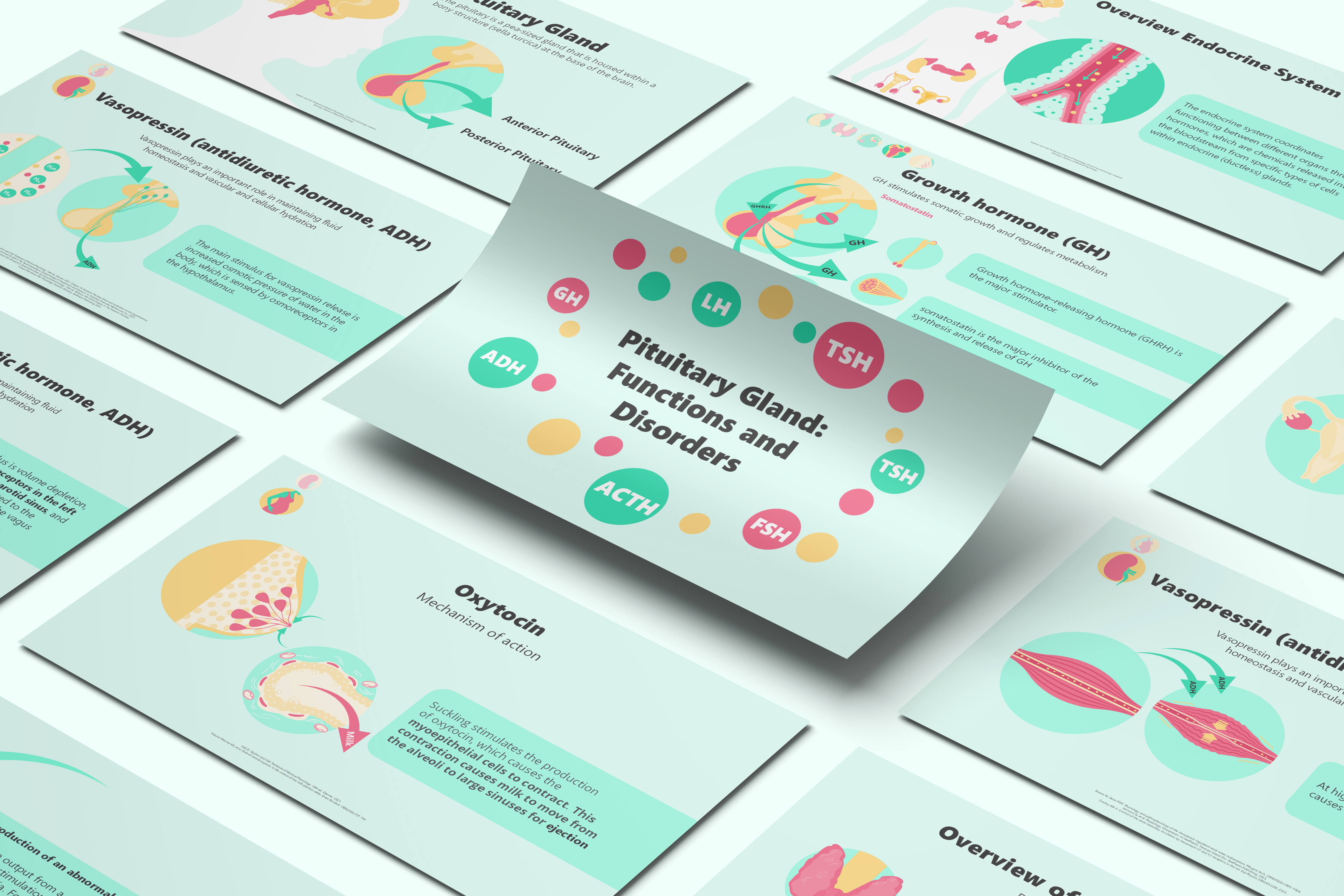
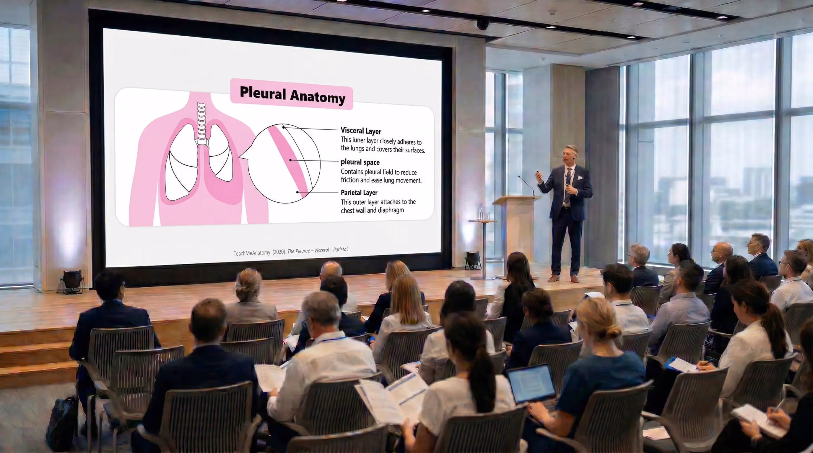
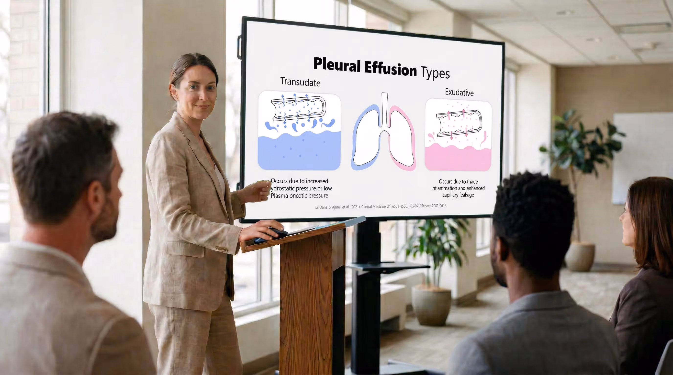
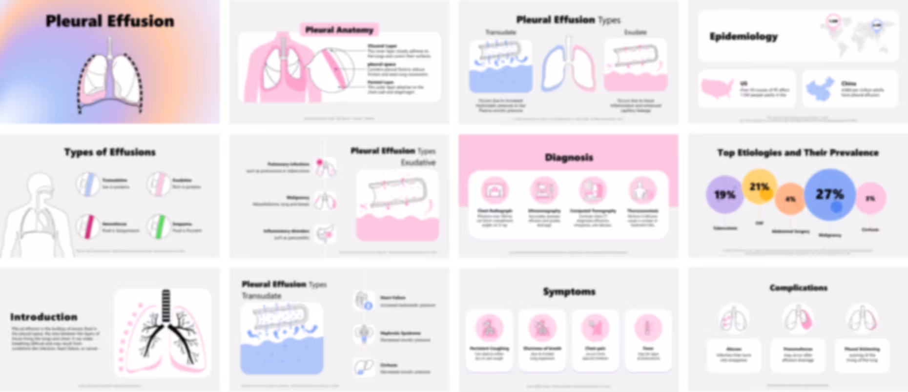
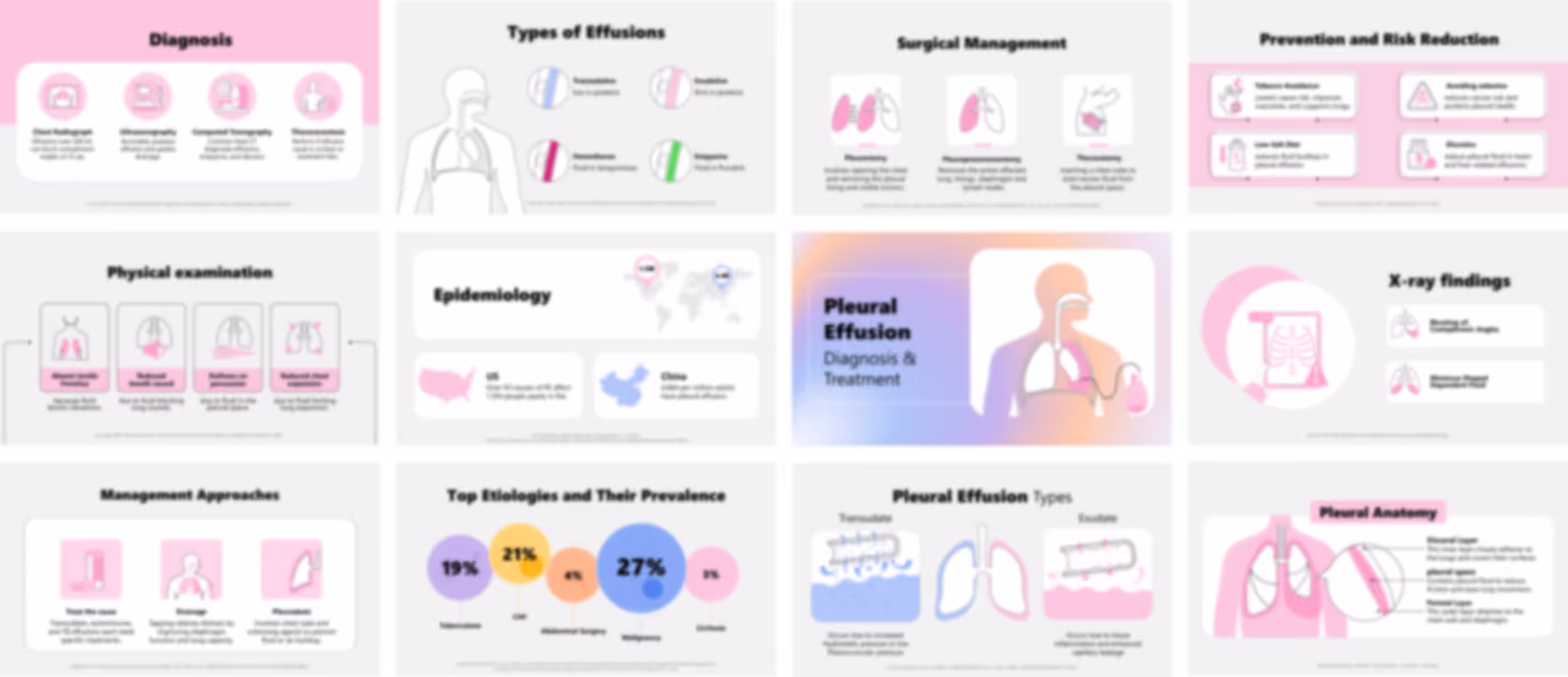
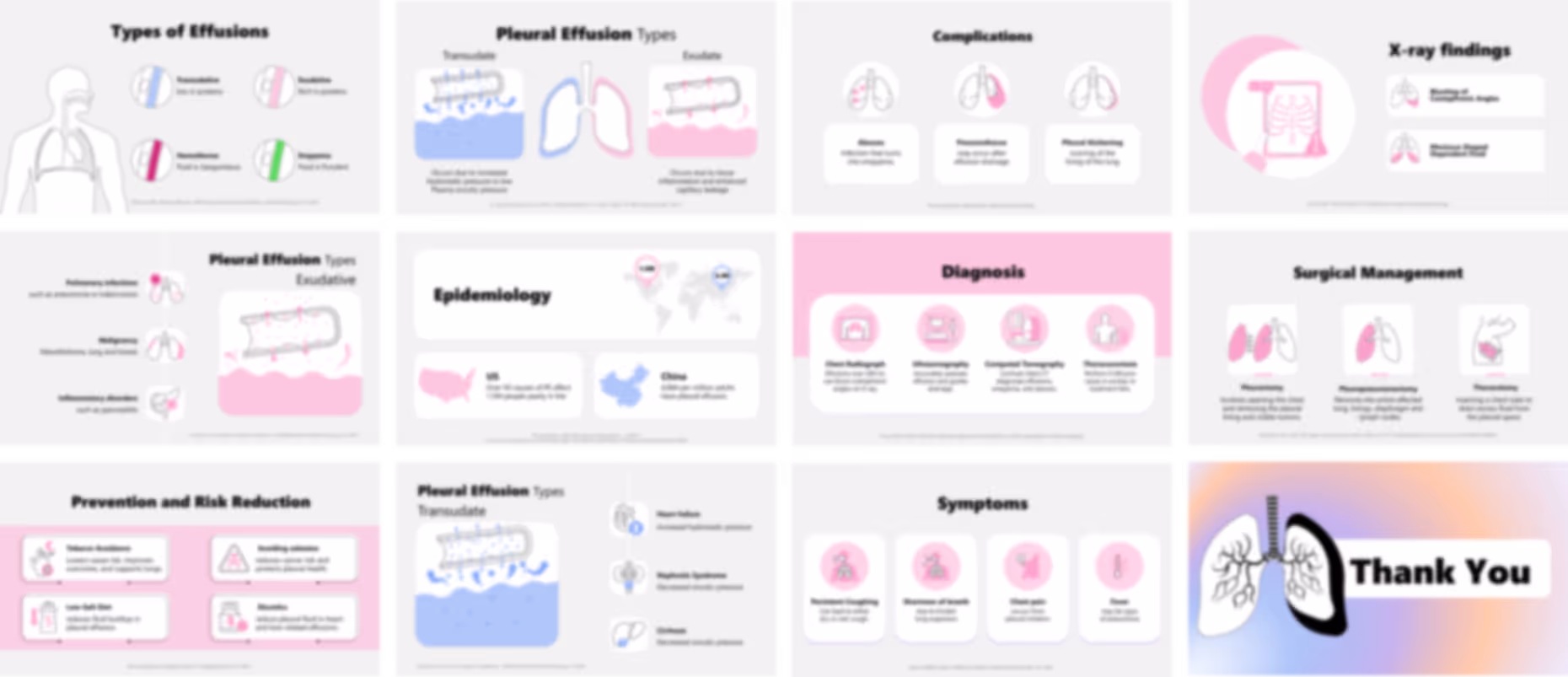
Pleural Effusion Presentation

No items found.
Trusted By Life Scinece Teams:

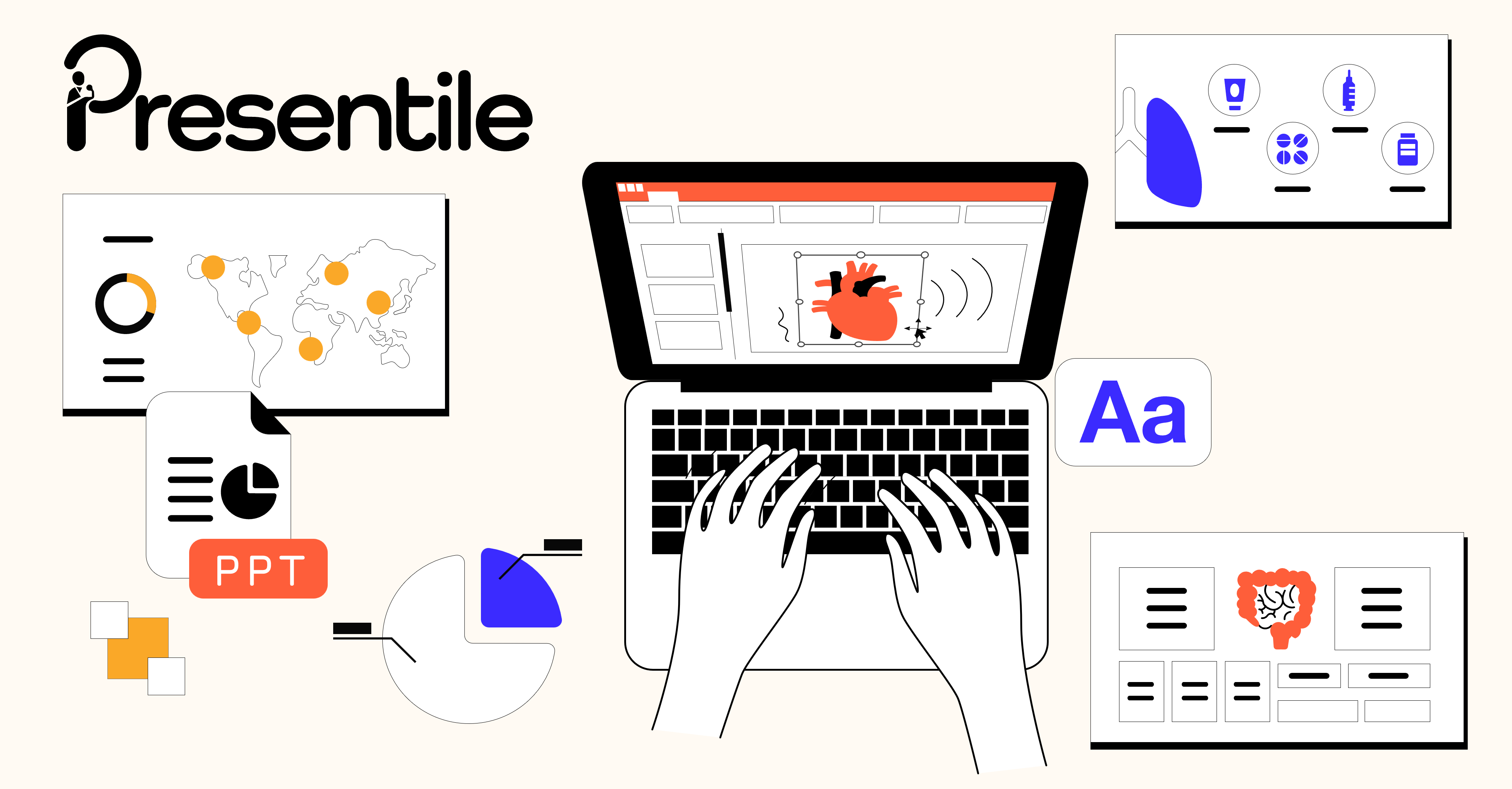
Fully editable in PowerPoint with customizable colors, shapes, and layouts
For easy and friendly drag and drop
Fully animated slides
To help simplify and communicate complex medical concepts
All graphics in vector format
For crisp visuals at any size
Medically referenced information and data
Built by medical professionals and researchers who perfectly understand the science behind the slides
Life sciences–focused design
Tailored for pharma, biotech, and healthcare organizations
Price:
$ 69.00 USD
For pharmaceutical marketing teams and medical affairs teams
- who need to communicate pleural effusion and respiratory information clearly and efficiently, this presentation provides a structured and professionally designed presentation deck that reduces preparation time, improves message clarity, and ensures consistent storytelling across educational materials and scientific discussions, with the flexibility to update and customize content based on your requirements.
For training managers, hospitals, and healthcare organizations

- who need to deliver structured respiratory education without developing content from scratch, this presentation offers a ready-to-use slide deck that simplifies complex clinical concepts and ensures consistent training delivery across workshops and internal programs.
For professors, academic lecturers, and universities
- who need clear and well-organized teaching materials, this presentation supports effective explanation of pleural anatomy, fluid mechanisms, and classification through a logical and expandable presentation deck that enhances student understanding and aligns with academic guidelines.
For healthcare professionals including pulmonologists, oncologists, radiologists, and internal medicine physicians
- who need to present clinical information clearly and efficiently, this presentation provides an organized slide deck that improves communication, supports structured delivery, and enhances engagement during conferences and clinical education sessions.
For researchers, biotech and medical device companies, and consultants
- who need to create structured scientific and strategic presentations, this presentation offers a flexible presentation deck that can be expanded and adapted for different audiences, projects, and communication goals.
No items found.